. 2011 Feb;193(3):599-610.
doi: 10.1128/JB.01203-10. Epub 2010 Dec 3.

Flavobacterium johnsoniae sprB is part of an operon spanning the additional gliding motility genes sprC, sprD, and sprF

Affiliations

Flavobacterium johnsoniae sprB is part of an operon spanning the additional gliding motility genes sprC, sprD, and sprF

Ryan G Rhodes et al. J Bacteriol. 2011 Feb.

Abstract

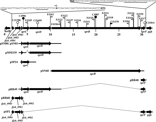
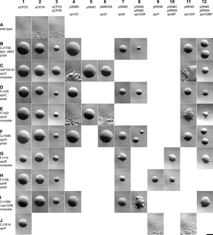
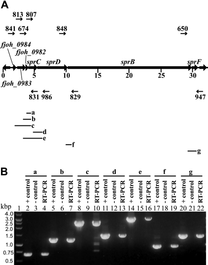
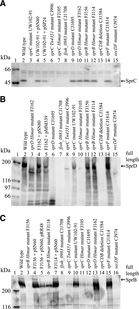
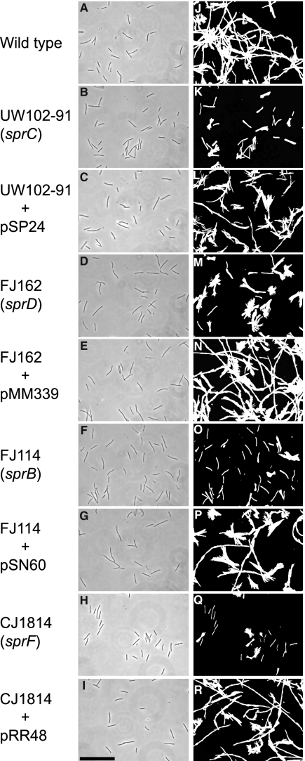
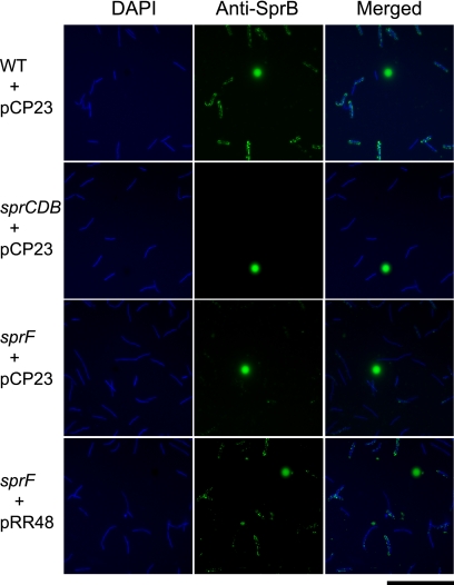
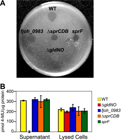
Cells of Flavobacterium johnsoniae move rapidly over surfaces by a process known as gliding motility. Gld proteins are thought to comprise the gliding motor that propels cell surface adhesins, such as the 669-kDa SprB. A novel protein secretion apparatus called the Por secretion system (PorSS) is required for assembly of SprB on the cell surface. Genetic and molecular analyses revealed that sprB is part of a seven-gene operon spanning 29.3 kbp of DNA. In addition to sprB, three other genes of this operon (sprC, sprD, and sprF) are involved in gliding. Mutations in sprB, sprC, sprD, and sprF resulted in cells that failed to form spreading colonies on agar but that exhibited some motility on glass in wet mounts. SprF exhibits some similarity to Porphyromonas gingivalis PorP, which is required for secretion of gingipain protease virulence factors via the P. gingivalis PorSS. F. johnsoniae sprF mutants produced SprB protein but were defective in localization of SprB to the cell surface, suggesting a role for SprF in secretion of SprB. The F. johnsoniae PorSS is involved in secretion of extracellular chitinase in addition to its role in secretion of SprB. SprF was not needed for chitinase secretion and may be specifically required for SprB secretion by the PorSS. Cells with nonpolar mutations in sprC or sprD produced and secreted SprB and propelled it rapidly along the cell surface. Multiple paralogs of sprB, sprC, sprD, and sprF are present in the genome, which may explain why mutations in sprB, sprC, sprD, and sprF do not result in complete loss of motility and suggests the possibility that semiredundant SprB-like adhesins may allow movement of cells over different surfaces.


Figures

FIG. 1.
FIG. 1.
Map of the sprB operon. Numbers below the map refer to kilobase pairs of the sequence. The sites of HimarEm1, HimarEm2, Tn4351, and pLYL03 insertions are indicated by filled triangles, open triangles, filled circles, and open circles, respectively. Orientations of HimarEm insertions are indicated by the direction in which the triangles are pointing. Triangles pointing to the right (FJ114, for example) have IR2 on the right side and the kanamycin resistance gene of the transposon reading toward the right, and they typically result in nonpolar mutations. The regions of DNA carried by plasmids used in this study are indicated beneath the map.
FIG. 2.
FIG. 2.
Photomicrographs of F. johnsoniae colonies. Colonies were grown for 48 h at 25°C on PY2 agar medium containing the appropriate antibiotics. Photomicrographs were taken with a Photometrics Cool-SNAPcf2 camera mounted on an Olympus IMT-2 phase-contrast microscope. Bar (at lower right; applies to all panels), 1 mm. Rows (A to J) indicate different strains of F. johnsoniae, and columns (1 to 12) indicate different plasmids introduced into these strains. Rows: A, wild-type F. johnsoniae UW101; B, fjoh_0983 insertion mutant CJ1708; C, sprC mutant UW102-91; D, sprC HimarEm2 insertion mutant FJ105; E, sprD HimarEm2 insertion mutant FJ162; F, sprD insertion mutant CJ1695; G, sprB HimarEm2 insertion mutant FJ114; H, sprB HimarEm2 insertion mutant FJ156; I, sprCDB deletion mutant CJ1584; J, sprF insertion mutant CJ1814. Columns: 1, control vector pCP23; 2, control vector pCP29; 3, control vectors pCP23 and pCP29; 4, pSN80 expressing sprC and sprD; 5, pSN81 carrying sprC and sprD in the opposite orientation as pSN80; 6, pMM339 expressing sprD; 7, pSN60 expressing sprB; 8, pSN80 (sprC sprD) and pSN60 (sprB); 9, pRR48 expressing sprF; 10, pRR48 (sprF) and pSN60 (sprB); 11, pRR49 expressing sprC, sprD, and sprF; 12, pRR49 (sprC sprD sprF) and pSN60 (sprB).
FIG. 3.
FIG. 3.
RT-PCR of the sprB operon. (A) Map of the sprB operon with primers used for RT-PCR (numbered arrows) and predicted PCR products (a to g; diagramed below in Fig. 3A). (B) RT-PCR of F. johnsoniae wild-type RNA to determine the sprB transcriptional unit. Antisense primer 831 and sense primers 674 (lane 4), 813 (lane 7), and 841 (lane 10) were used to amplify across the junctions spanning fjoh_0984 to sprC. Antisense primer 986 and sense primers 807 (lane 13) and 813 (lane 16) were used to amplify across the junctions spanning fjoh_0983 to sprD. Antisense primer 829 was used with sense primer 848 to amplify across the sprD-sprB junction (lane 19), and antisense primer 947 was used with sense primer 650 to amplify across the sprB-sprF junction (lane 22). For each primer pair, three reaction mixtures were loaded on the gel: a positive control PCR using F. johnsoniae chromosomal DNA as template (lanes 2, 5, 8, 11, 14, 17, and 20), a no-RT control reaction mixture (lanes 3, 6, 9, 12, 15, 18, and 21), and a RT-PCR mixture. A 1-kb ladder was used as a size standard (lane 1).
FIG. 4.
FIG. 4.
Western immunoblot detection of SprC, SprD, and SprB in whole-cell extracts of wild-type and mutant strains of F. johnsoniae. (A) Detection of SprC. Lane 1, molecular mass markers; lane 2, wild-type F. johnsoniae UW101; lane 3, sprC point mutant UW102-91; lane 4, UW102-91 complemented with pSN80; lane 5, UW102-91 with pSP24; lane 6, sprC Tn4351 insertion mutant CJ996; lane 7, sprC HimarEm2 insertion mutant FJ105; lane 8, fjoh_0983 insertion mutant CJ1708; lane 9, sprD insertion mutant CJ1695; lane 10, sprD HimarEm2 insertion mutant FJ162; lane 11, sprB HimarEm2 insertion mutant FJ156; lane 12, sprB HimarEm2 insertion mutant FJ114; lane 13, sprCDB deletion mutant CJ1584; lane 14, sprF insertion mutant CJ1814; lane 15, secDF mutant CJ974. (B) Detection of SprD. Lane 1, molecular mass markers; lane 2, wild-type F. johnsoniae; lane 3, sprD HimarEm2 insertion mutant FJ162; lane 4, FJ162 complemented with pSN80; lane 5, FJ162 complemented with pMM339; lane 6, sprD insertion mutant CJ1695; lane 7, fjoh_0983 insertion mutant CJ1708; lane 8, sprC Tn4351 insertion mutant CJ996; lane 9, sprC point mutant UW102-91; lane 10, sprC HimarEm2 insertion mutant FJ105; lane 11, sprB HimarEm2 insertion mutant FJ156; lane 12, sprB HimarEm2 insertion mutant FJ114; lane 13, sprCDB deletion mutant CJ1584; lane 14, sprF insertion mutant CJ1814; lane 15, secDF mutant CJ974. (C) Detection of SprB. Lane 1, molecular mass markers; lane 2, wild-type F. johnsoniae; lane 3, sprB HimarEm2 insertion mutant FJ156; lane 4, FJ156 with pSN60; lane 5, FJ156 complemented with pSN60 and pRR48; lane 6, sprB HimarEm2 insertion mutant FJ114; lane 7, FJ114 with pSN60; lane 8, fjoh_0983 insertion mutant CJ1708; lane 9, sprC Tn4351 insertion mutant CJ996; lane 10, sprC point mutant UW102-91; lane 11, sprC HimarEm2 insertion mutant FJ105; lane 12, sprD insertion mutant CJ1695; lane 13, sprD HimarEm2 insertion mutant FJ162; lane 14, sprCDB deletion mutant CJ1584; lane 15, sprF insertion mutant CJ1814; lane 16, secDF mutant CJ974. Forty micrograms of protein was loaded in each lane. Some strains carried empty control vector pCP23 (panel A, lanes 2, 3, 6, 7, 8, 9, 10, 11, 13, and 14; panel B, lanes 2, 3, 6, 7, 8, 9, 10, 11, 13, and 14; panel C, lanes 2, 8, 9, 10, 11, 12, 13, 14, and 15) or pCP29 (panel A, lane 12; panel B, lanes 8, 10, and 12; panel C, lanes 3 and 6), which had no effect on expression of SprB, SprC, or SprD.
FIG. 5.
FIG. 5.
Effects of mutations in sprC, sprD, sprB, and sprF on gliding of cells on glass. Cells attached to a glass coverslip on a Palmer cell were observed by phase-contrast microscopy, and digital images of cells of the wild-type strain with control vector pCP23 (A), sprC mutant UW102-91 with pCP23 (B), UW102-91 complemented with pSP24 (C), sprD mutant FJ162 with pCP23 (D), FJ162 complemented with pMM339 (E), sprB mutant FJ114 with control vector pCP29 (F), FJ114 complemented with pSN60 (G), sprF mutant CJ1814 with pCP23 (H), and CJ1814 complemented with pRR48 (I) were recorded at time zero. Tracks illustrating the movements of the cells shown in panels A to I over a 60-s period were obtained by superimposing individual digital video frames of the wild-type strain with pCP23 (J), sprC mutant UW102-91 with pCP23 (K), UW102-91 complemented with pSP24 (L), sprD mutant FJ162 with pCP23 (M), FJ162 complemented with pMM339 (N), sprB mutant FJ114 with pCP29 (O), FJ114 complemented with pSN60 (P), sprF mutant CJ1814 with pCP23 (Q), and CJ1814 complemented with pRR48 (R). Images were recorded using a Photometrics CoolSNAPcf2 camera mounted on an Olympus BH-2 phase-contrast microscope. Bar (shown in panel I; applies to all panels), 40 μm.
FIG. 6.
FIG. 6.
Detection of surface-localized SprB protein by immunofluorescence microscopy. Cells of wild-type and mutant F. johnsoniae were exposed to DAPI and to anti-SprB antibodies followed by secondary antibodies conjugated to Alexa-488. The fluorescent spheres are InSpeck relative intensity control fluorescent beads. WT, wild-type F. johnsoniae UW101; sprCDB, sprCDB deletion mutant CJ1584; sprF, sprF insertion mutant CJ1814. pCP23 is a control vector; pRR48 carries sprF. Bar, 20 μm.
FIG. 7.
FIG. 7.
Mutations in genes of the sprCDBF operon have no effect on chitin utilization. (A) Digestion of chitin by intact cells. Approximately 106 cells of F. johnsoniae strains were spotted on MYA-chitin medium and incubated at 25°C for 4 days. Strains included wild-type F. johnsoniae UW101 (WT), fjoh_0983 insertion mutant CJ1708 (fjoh_0983), sprCDB deletion strain CJ1584 (ΔsprCDB), sprF insertion mutant CJ1814 (sprF), and nonmotile gldNO deletion mutant CJ1631A (ΔgldNO). Erythromycin was included in the medium to ensure maintenance of the plasmid insertions in fjoh_0983 and sprF. The wild type, CJ1584, and CJ1631A carried control vector pCP29, which conferred erythromycin resistance and allowed these strains to be tested on the same medium as the fjoh_0983 and sprF insertion mutants. Control experiments verified that pCP29 had no effect on chitin utilization. (B) Chitinase activities of cell lysates and culture supernatants of F. johnsoniae strains, determined using the synthetic substrate 4-MU-(GlcNAc)2. Equal amounts of each sample, based on the protein content of the original cell suspension, were incubated with 10 nmol of 4-MU-(GlcNAc)2 for 4 h at 37°C, and the amount of 4-MU released was determined by measuring fluorescence emission at 460 nm following excitation at 360 nm. WT, F. johnsoniae UW101; ΔgldNO, gldNO deletion mutant CJ1631A; fjoh_0983, fjoh_0983 insertion mutant CJ1708; ΔsprCDB, sprCDB deletion mutant CJ1584; sprF, sprF insertion mutant CJ1814. All strains in panel B carried control vector pCP23, which has no effect on chitinase activity.
FIG. 8.
FIG. 8.
Effects of mutations on bacteriophage resistance. Bacteriophages (in 5-μl aliquots of lysates containing approximately 109 PFU/ml) were spotted onto lawns of cells in CYE overlay agar. The plates were incubated at 25°C for 24 h to observe lysis. Bacteriophages were spotted in the following order from left to right, as indicated also by the numbers in panel A: top row, φCj1, φCj13, and φCj23; middle row, φCj28, φCj29, and φCj42; bottom row, φCj48 and φCj54. (A) Wild-type F. johnsoniae UW101 with control vector pCP23; (B) fjoh_0983 mutant CJ1708 with pCP23; (C) sprC mutant FJ105 with pCP23; (D) sprD mutant CJ1695 with pCP23; (E) sprB mutant FJ114 with control vector pCP29; (F) sprB mutant FJ114 with pSN60, which carries sprB; (G) sprF mutant CJ1814 with pCP23; (H) sprF mutant CJ1814 with pRR48, which carries sprF.

Similar articles

Cited by

References

    1. Agarwal, S., D. W. Hunnicutt, and M. J. McBride. 1997. Cloning and characterization of the Flavobacterium johnsoniae (Cytophaga johnsonae) gliding motility gene, gldA. Proc. Natl. Acad. Sci. U. S. A. 94:12139-12144. - PMC - PubMed
    1. Altschul, S. F., W. Gish, W. Miller, E. W. Myers, and D. J. Lipman. 1990. Basic local alignment search tool. J. Mol. Biol. 215:403-410. - PubMed
    1. Alvarez, B., P. Secades, M. J. McBride, and J. A. Guijarro. 2004. Development of genetic techniques for the psychrotrophic fish pathogen Flavobacterium psychrophilum. Appl. Environ. Microbiol. 70:581-587. - PMC - PubMed
    1. Bendtsen, J. D., H. Nielsen, G. von Heijne, and S. Brunak. 2004. Improved prediction of signal peptides: SignalP 3.0. J. Mol. Biol. 340:783-795. - PubMed
    1. Braun, T. F., M. K. Khubbar, D. A. Saffarini, and M. J. McBride. 2005. Flavobacterium johnsoniae gliding motility genes identified by mariner mutagenesis. J. Bacteriol. 187:6943-6952. - PMC - PubMed

Publication types

Substances

LinkOut - more resources