. 2011 Mar;120(1):68-78.
doi: 10.1093/toxsci/kfq360. Epub 2010 Dec 3.

Aryl hydrocarbon receptor activation by TCDD reduces inflammation associated with Crohn's disease

Affiliations

Aryl hydrocarbon receptor activation by TCDD reduces inflammation associated with Crohn's disease

Jenna M Benson et al. Toxicol Sci. 2011 Mar.

Abstract

Crohn's disease results from a combination of genetic and environmental factors that trigger an inappropriate immune response to commensal gut bacteria. The aryl hydrocarbon receptor (AhR) is well known for its involvement in the toxicity of 2,3,7,8-tetrachlorodibenzo-p-dioxin (TCDD), an environmental contaminant that affects people primarily through the diet. Recently, TCDD was shown to suppress immune responses by generating regulatory T cells (Tregs). We hypothesized that AhR activation dampens inflammation associated with Crohn's disease. To test this hypothesis, we utilized the 2,4,6-trinitrobenzenesulfonic acid (TNBS) murine model of colitis. Mice were gavaged with TCDD prior to colitis induction with TNBS. Several parameters were examined including colonic inflammation via histological and flow cytometric analyses. TCDD-treated mice recovered body weight faster and experienced significantly less colonic damage. Reduced levels of interleukin (IL) 6, IL-12, interferon-gamma, and tumor necrosis factor-α demonstrated suppression of inflammation in the gut following TCDD exposure. Forkhead box P3 (Foxp3)(egfp) mice revealed that TCDD increased the Foxp3+ Treg population in gut immune tissue following TNBS exposure. Collectively, these results suggest that activation of the AhR by TCDD decreases colonic inflammation in a murine model of colitis in part by generating regulatory immune cells. Ultimately, this work may lead to the development of more effective therapeutics for the treatment of Crohn's disease.


Figures

FIG. 1.
FIG. 1.
This schematic represents the experimental design utilized for the studies in which animals were gavaged with 15 μg/kg TCDD, peripherally sensitized with 5% TNBS, intrarectally injected with 2.5 mg TNBS, and subsequently harvested for evaluation.
FIG. 2.
FIG. 2.
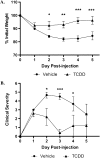
TCDD reduces wasting disease and clinical symptoms associated with TNBS-induced colitis. Animals were monitored daily after disease induction for body weight loss (A) and other clinical symptoms including body condition, dehydration state, and stool consistency (B). Clinical severity was determined by combining the symptom scores for a total possible severity score of nine, as described in Supplementary table 1A. Results are shown as mean ± SEM (n = 6) and representative of three separate experiments. “*” Indicates significances of p ≤ 0.05, “**” indicates significance of p ≤ 0.01, and “***” indicates significance of p ≤ 0.0001.
FIG. 3.
FIG. 3.
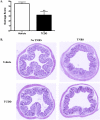
Colonic inflammation is reduced following exposure to TCDD. Animals were harvested 5 days after disease induction and sections of the distal colon were assessed microscopically. Damage score (A) was determined by combining the scores for inflammatory cell infiltration (0–3) and tissue damage (0–3), as described in Supplementary table 1B. Representative images of H&E-stained tissue sections of vehicle-treated and TCDD-treated animals are shown in (B). Results are shown as mean ± SEM (n = 6) and are representative of three separate experiments. “**” Indicates significance of p ≤ 0.01.
FIG. 4.
FIG. 4.
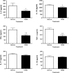
Modulation of cytokine production by TCDD in colon tissue. On day 5 following disease induction, colons were excised, washed, and homogenized. The supernatants were assessed for cytokine production, as described in the “Materials and Methods” section. Results are shown as mean ± SEM (n = 6) and representative of three separate experiments. “*” Indicates significance of p ≤ 0.05 and “**” indicates significance of p ≤ 0.01.
FIG. 5.
FIG. 5.
TCDD increases Foxp3 expression in colon tissue. Colons were harvested from Foxp3egfp mice 3 days after disease induction. Tissues were sectioned, mounted on slides, and stained with 4′,6–diamidino–2–phenylindole to visualize the nucleus. Slides were imaged at ×40 magnification using the Olympus FluoView 1000 LSC on an inverted 1 × 81 microscope with spectral detection and total internal reflection fluorescence module. Images are representative of a single experiment (n = 6).

Similar articles

Cited by

References

    1. Abraham C, Cho JH. IL-23 and autoimmunity: new insights into the pathogenesis of inflammatory bowel disease. Annu. Rev. Med. 2009;60:97–110. - PubMed
    1. Aggarwal BB, Shishodia S. Molecular targets of dietary agents for prevention and therapy of cancer. Biochem. Pharmacol. 2006;71:1397–1421. - PubMed
    1. Bankoti J, Burnett A, Navarro S, Miller AK, Rase B, Shepherd DM. Effects of TCDD on the fate of naive dendritic cells. Toxicol. Sci. 2010a;115:422–434. - PMC - PubMed
    1. Bankoti J, Rase B, Simones T, Shepherd DM. Functional and phenotypic effects of AhR activation in inflammatory dendritic cells. Toxicol. Appl. Pharmacol. 2010b;246:18–28. - PMC - PubMed
    1. Baumgart DC, Dignass AU. Intestinal barrier function. Curr. Opin. Clin. Nutr. Metab. Care. 2002;5:685–694. - PubMed

Publication types

MeSH terms