

Polyethyleneimine nanoparticles incorporated into resin composite cause cell death and trigger biofilm stress in vivo
- PMID: 21131569
- PMCID: PMC3009770
- DOI: 10.1073/pnas.1010341107
Polyethyleneimine nanoparticles incorporated into resin composite cause cell death and trigger biofilm stress in vivo
Abstract
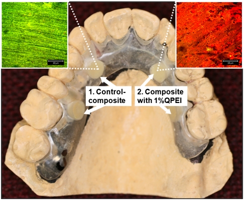
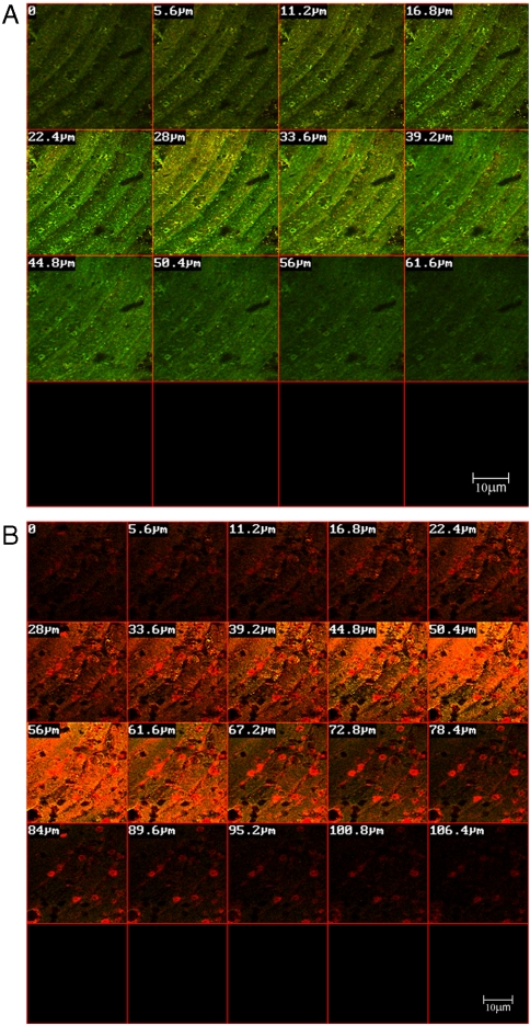
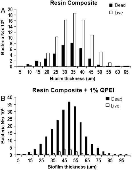
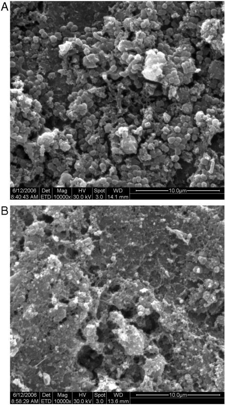
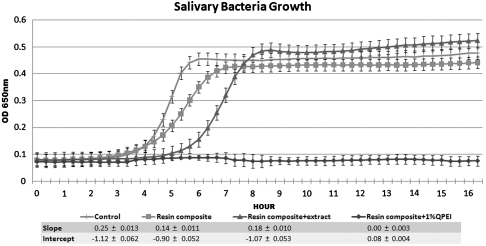
Incorporation of cross-linked quaternary ammonium polyethylenimine (QPEI) nanoparticles in dental resin composite has a long-lasting and wide antimicrobial effect with no measured impact on biocompatibility in vitro. We hypothesized that QPEI nanoparticles incorporated into a resin composite have a potent antibacterial effect in vivo and that this stress condition triggers a suicide module in the bacterial biofilm. Ten volunteers wore a removable acrylic appliance, in which two control resin composite specimens and two resin composite specimens incorporating 1% wt/wt QPEI nanoparticles were inserted to allow the buildup of intraoral biofilms. After 4 h, the specimens were removed and tested for bacterial vitality and biofilm thickness, using confocal laser scanning microscopy. The vitality rate in specimens incorporating QPEI was reduced by > 50% (p < 0.00001), whereas biofilm thickness was increased (p < 0.05). The ability of the biofilm supernatant to restore bacterial death was tested in vitro. The in vitro tests showed a 70% decrease in viable bacteria (p < 0.05). Biofilm morphological differences were also observed in the scanning electron microscope micrographs of the resin composite versus the resin composite incorporating QPEI. These results strongly suggest that QPEI nanoparticles incorporated at a low concentration in resin composite exert a significant in vivo antibiofilm activity and exhibit a potent broad spectrum antibacterial activity against salivary bacteria.
Conflict of interest statement
The authors declare no conflict of interest.
Figures
Similar articles
-
Characterisation of the antibacterial effect of polyethyleneimine nanoparticles in relation to particle distribution in resin composite.J Dent. 2015 Feb;43(2):287-94. doi: 10.1016/j.jdent.2014.05.003. Epub 2014 May 29. J Dent. 2015. PMID: 24881908
-
Lethal bacterial trap: Cationic surface for endodontic sealing.J Biomed Mater Res A. 2016 Feb;104(2):427-34. doi: 10.1002/jbm.a.35576. Epub 2015 Oct 20. J Biomed Mater Res A. 2016. PMID: 26418438
-
Antibacterial effect of composite resin foundation material incorporating quaternary ammonium polyethyleneimine nanoparticles.J Prosthet Dent. 2016 Oct;116(4):603-609. doi: 10.1016/j.prosdent.2016.02.022. Epub 2016 May 5. J Prosthet Dent. 2016. PMID: 27157602
-
Application of Antimicrobial Polymers in the Development of Dental Resin Composite.Molecules. 2020 Oct 15;25(20):4738. doi: 10.3390/molecules25204738. Molecules. 2020. PMID: 33076515 Free PMC article. Review.
-
The germicidal effect, biosafety and mechanical properties of antibacterial resin composite in cavity filling.Heliyon. 2023 Aug 22;9(9):e19078. doi: 10.1016/j.heliyon.2023.e19078. eCollection 2023 Sep. Heliyon. 2023. PMID: 37662807 Free PMC article. Review.
Cited by
-
Polyethylenimine Increases Antibacterial Efficiency of Chlorophyllin.Antibiotics (Basel). 2022 Oct 7;11(10):1371. doi: 10.3390/antibiotics11101371. Antibiotics (Basel). 2022. PMID: 36290029 Free PMC article.
-
Sustained Release of Antibacterial Lipopeptides from Biodegradable Polymers against Oral Pathogens.PLoS One. 2016 Sep 8;11(9):e0162537. doi: 10.1371/journal.pone.0162537. eCollection 2016. PLoS One. 2016. PMID: 27606830 Free PMC article.
-
Recent Advances in Strategies to Combat Bacterial Drug Resistance: Antimicrobial Materials and Drug Delivery Systems.Pharmaceutics. 2023 Apr 7;15(4):1188. doi: 10.3390/pharmaceutics15041188. Pharmaceutics. 2023. PMID: 37111673 Free PMC article. Review.
-
Quaternary Ammonium Silica Nanoparticles for Antimicrobial Implantable Medical Devices: An In Vitro Study.Life (Basel). 2024 Dec 12;14(12):1654. doi: 10.3390/life14121654. Life (Basel). 2024. PMID: 39768361 Free PMC article.
-
Novel dental composite with capability to suppress cariogenic species and promote non-cariogenic species in oral biofilms.Mater Sci Eng C Mater Biol Appl. 2019 Jan 1;94:587-596. doi: 10.1016/j.msec.2018.10.004. Epub 2018 Oct 2. Mater Sci Eng C Mater Biol Appl. 2019. PMID: 30423744 Free PMC article.
References
-
- Hahn R, Weiger R, Netuschil L, Bruch M. Microbial accumulation and vitality on different restorative materials. Dent Mater. 1993;9:312–316. - PubMed
-
- Siegrist BE, Brecx MC, Gusberti FA, Joss A, Lang NP. In vivo early human dental plaque formation on different supporting substances. A scanning electron microscopic and bacteriological study. Clin Oral Implan Res. 1991;2:38–46. - PubMed
-
- Chigira H, et al. Efficacy of various commercial dentin bonding systems. Dent Mater. 1994;10:363–368. - PubMed
-
- Goracci G, Mori G, Bazzucchi M. Marginal seal and biocompatibility of a fourth-generation bonding agent. Dent Mater. 1995;11:343–347. - PubMed
-
- Lobo MM, Goncalves RB, Ambrosano GM, Pimenta LA. Chemical or microbiological models of secondary caries development around different dental restorative materials. J Biomed Mater Res B. 2005;74:725–731. - PubMed
Publication types
MeSH terms
Substances
LinkOut - more resources
Full Text Sources
Other Literature Sources
Medical
