. 2011 Jan 19;30(2):277-88.
doi: 10.1038/emboj.2010.310. Epub 2010 Dec 3.

TDP-43 regulates its mRNA levels through a negative feedback loop

Affiliations

TDP-43 regulates its mRNA levels through a negative feedback loop

Youhna M Ayala et al. EMBO J. .

Abstract

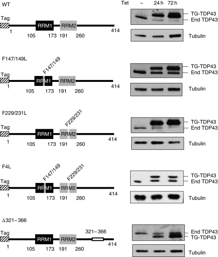
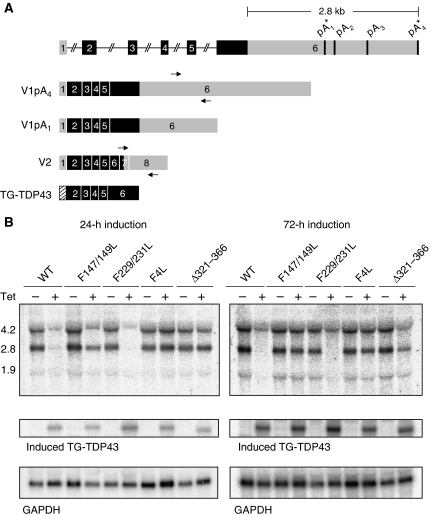
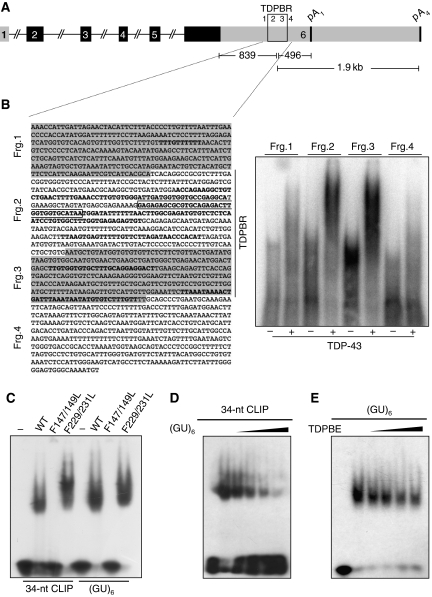
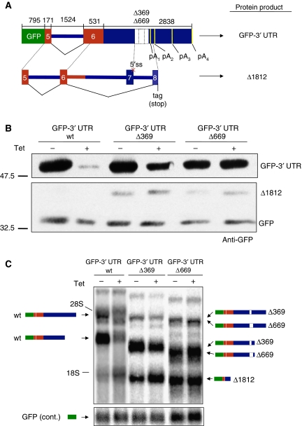
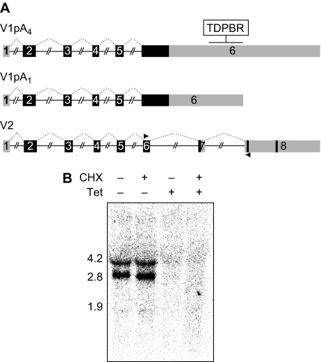
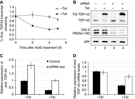
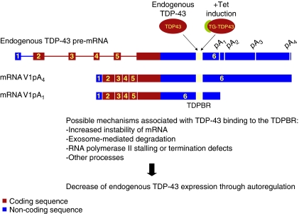
TAR DNA-binding protein (TDP-43) is an evolutionarily conserved heterogeneous nuclear ribonucleoprotein (hnRNP) involved in RNA processing, whose abnormal cellular distribution and post-translational modification are key markers of certain neurodegenerative diseases, such as amyotrophic lateral sclerosis and frontotemporal lobar degeneration. We generated human cell lines expressing tagged forms of wild-type and mutant TDP-43 and observed that TDP-43 controls its own expression through a negative feedback loop. The RNA-binding properties of TDP-43 are essential for the autoregulatory activity through binding to 3' UTR sequences in its own mRNA. Our analysis indicated that the C-terminal region of TDP-43, which mediates TDP-43-hnRNP interactions, is also required for self-regulation. TDP-43 binding to its 3' UTR does not significantly change the pre-mRNA splicing pattern but promotes RNA instability. Moreover, blocking exosome-mediated degradation partially recovers TDP-43 levels. Our findings demonstrate that cellular TDP-43 levels are under tight control and it is likely that disease-associated TDP-43 aggregates disrupt TDP-43 self-regulation, thus contributing to pathogenesis.


Conflict of interest statement

The authors declare that they have no conflict of interest.

Figures

Figure 1
Figure 1
Induced expression of wild-type and mutant TDP-43 in HEK293 cells. Tagged forms of TDP-43 (F-TDP43) were expressed for 24 or 72 h on tetracycline (Tet) induction (1 μg/ml). The diagrams on the left are a schematic representation of the different TDP-43 protein domains, numbering denotes amino acid residues. The N-terminal domain tag is represented by the hashed box and the RNA recognition motifs 1 and 2 are shown as black and grey boxes, respectively. Deletion of the C-terminal region in mutant Δ321–366 is depicted as a white box. The right panels are immunoblot analyses of the protein extracts with an antibody against TDP-43 that detects both endogenous (end) and tagged (TG-TDP43) protein. Tubulin was used as loading control.
Figure 2
Figure 2
TDP-43 negative autogenous regulation occurs at the transcript level. (A) Schematic representation of the TDP-43 gene with coding exons and untranslated regions shown in black and grey, respectively. The various predicted polyadenylation signals are indicated, where (*) denotes the experimentally validated sites. The two principally expressed endogenous isoforms (V1pA1 and V1pA4) are shown including the transgenic TG-TDP43 transcript containing the coding sequences only. The V2 form corresponds to a minor isoform discussed in Supplementary Figure 2. (B) Northern blot analysis of control and after 24 and 72 h of induction. V1pA1 and V1pA4 correspond to the 2.8 and 4.2 Kb bands, respectively. Endogenous TDP-43 transcripts were detected by probing the region indicated by the arrows in (A), while TG-TDP43 was identified with a probe spanning exons 2 and 3. GAPDH detection was used as loading control.
Figure 3
Figure 3
TDP-43 specifically binds its 3′ UTR region. (A) Schematic representation of the TDP-43 gene depicting the location and sequence of the TDPBR (B, fragments 2 and 3) within the 3′ UTR. Coding exons and untranslated regions are shown in black and grey, respectively. (B) In the left panel, the 3′ UTR region of TDP-43 with the different regions tested for TDP-43 binding (frgs. 1–4) is reported. In this panel, all the CLIP sequences/clusters reported from different experiments (J Ule and J Tollervey unpublished results) are highlighted in bold. The major 34-nt CLIP sequence seen in HEK 293 cells and used in comparative-binding analysis (CE) is boxed. On the right, band-shift analysis using recombinant TDP-43 using these different fragments. (C) GST-TDP43 mutants were analyzed for binding to the major 34-nt CLIP sequence (boxed) and (GU)6 RNA as control. (D, E) Competition assays using constant levels of wild-type protein to bind radiolabelled (GU)6 as a function of increasing concentrations of unlabelled 34-nt CLIP sequence (D), or binding to radiolabelled 34-nt CLIP sequence as a function of increasing concentrations unlabelled (GU)6. Protein was absent from the first lane in each panel. ∼0.5 μg of the different recombinant proteins were used with 1 ng of the various 5′-labeled single-stranded RNA oligonucleotides. Competition analyses were carried out with 2.5, 3.75, 5, and 10 ng of unlabelled 34-nt CLIP sequence and (GU)6.
Figure 4
Figure 4
A region in the 3′ UTR of TDP-43 mediates autoregulation (A) a schematic diagram of the GFP-3′ UTR wt and Δ369 and Δ669 constructs. Wild-type TDP-43 coding sequences are shown in grey. The lower panel shows the splicing events that lead to the Δ1812 mRNA and protein isoforms. (B) GFP protein expression when the GFP-3′ UTR wt and Δ369 and Δ669 constructs were transfected into the HEK293 stable cell lines following induction (+) of the TG-TDP43 transgene. Transfection efficiencies were normalized by co-transfecting a GFP expression vector (lower panel). In the GFP-3′ UTR Δ369 and Δ669 (−) and (+) lanes, the western blot also contains an additional 40 kDa protein band that was called Δ1812. The different proteins were detected with anti-GFP (C) shows a northern blot analysis of the transcripts derived from the GFP-3′ UTR wt, Δ369, and Δ669 constructs in the HEK293 stable cell lines before (−) and after Tet induction (+). All constructs produce two major mRNA species because of the usage of different polyadenylation sites. In addition to these species, the GFP-3′ UTR Δ369 and Δ669 constructs also produce an aberrantly spliced mRNA isoform (Δ1812) that accounts for the production of the aberrant protein present in the western blots (B).
Figure 5
Figure 5
TDP-43 autoregulation is independent of NMD. (A) The existence of alternative splicing that activates the removal of two downstream introns (V2) was seen in the EST database. The shorter form V2 was seen to undergo NMD (Supplementary Figure 2), while northern blot analysis (B) of control and Tet-induced samples in the presence and absence of CHX treatment (50 μg/ml, for 3 h) did not detect the presence of V2. V2 should have a MW of 1.2 or 2.5 Kb depending on the poly A site usage. The probe was generated with the primers shown in (A) followed by purification of the product corresponding to V2.
Figure 6
Figure 6
TDP-43 overexpression promotes RNA instability in an exosome-dependent fashion. (A) Transcript half-life was estimated after ActD (5 μg/ml) treatment of HEK293 control and induced cells. Transcript levels at the indicated times were analyzed by qPCR. The relative levels of expression at the different time points were normalized according to the levels at time 0, (−) and (+) Tet induction (empty and filled circles, respectively). (B) Exosome components, PM/Scl-100 and DIS3, were simultaneously silenced in control and induced cells and their effect on TDP-43 expression levels was observed in western blot. (C) Quantification of endogenous TDP-43 protein levels under the different conditions was normalized against p84 levels. Error bars, s.e.m.; n=3 experiments for both panels. (D) mRNA expression levels of endogenous TDP-43 mRNA levels in −Tet and +Tet conditions. Both the transcript levels were normalized to the GAPDH mRNA levels and determined by real-time qPCR. The mRNA levels were quantified separately in triplicates and s.d. values obtained are shown on the bars.
Figure 7
Figure 7
Regulation of TDP-43 expression model. Endogenous or transgenic TDP-43 (TG-TDP43) protein binds to the TDPBR found in the 3′ UTR of TDP-43 transcripts. This TDP-43–RNA interaction promotes, along with other possible events, RNA instability and degradation resulting in lower levels of TDP-43 gene expression.

References

    1. Amador-Ortiz C, Lin WL, Ahmed Z, Personett D, Davies P, Duara R, Graff-Radford NR, Hutton ML, Dickson DW (2007) TDP-43 immunoreactivity in hippocampal sclerosis and Alzheimer's disease. Ann Neurol 61: 435–445 - PMC - PubMed
    1. Arai T, Hasegawa M, Akiyama H, Ikeda K, Nonaka T, Mori H, Mann D, Tsuchiya K, Yoshida M, Hashizume Y, Oda T (2006) TDP-43 is a component of ubiquitin-positive tau-negative inclusions in frontotemporal lobar degeneration and amyotrophic lateral sclerosis. Biochem Biophys Res Commun 351: 602–611 - PubMed
    1. Ash PE, Zhang YJ, Roberts CM, Saldi T, Hutter H, Buratti E, Petrucelli L, Link CD (2010) Neurotoxic effects of TDP-43 overexpression in C. elegans. Hum Mol Genet 19: 3206–3218 - PMC - PubMed
    1. Ayala YM, Pantano S, D'Ambrogio A, Buratti E, Brindisi A, Marchetti C, Romano M, Baralle FE (2005) Human, Drosophila, and C. elegans TDP43: nucleic acid binding properties and splicing regulatory function. J Mol Biol 348: 575–588 - PubMed
    1. Buratti E, Baralle FE (2001) Characterization and functional implications of the RNA binding properties of nuclear factor TDP-43, a novel splicing regulator of CFTR exon 9. J Biol Chem 276: 36337–36343 - PubMed

Publication types