. 2011 Jan 19;30(2):341-54.
doi: 10.1038/emboj.2010.317. Epub 2010 Dec 3.

Dma1 ubiquitinates the SIN scaffold, Sid4, to impede the mitotic localization of Plo1 kinase

Affiliations

Dma1 ubiquitinates the SIN scaffold, Sid4, to impede the mitotic localization of Plo1 kinase

Alyssa E Johnson et al. EMBO J. .

Abstract

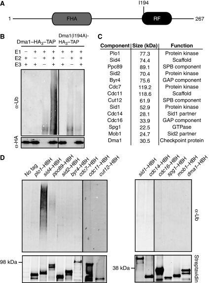
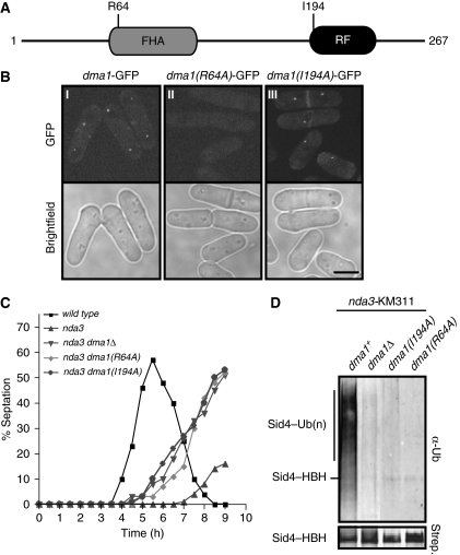
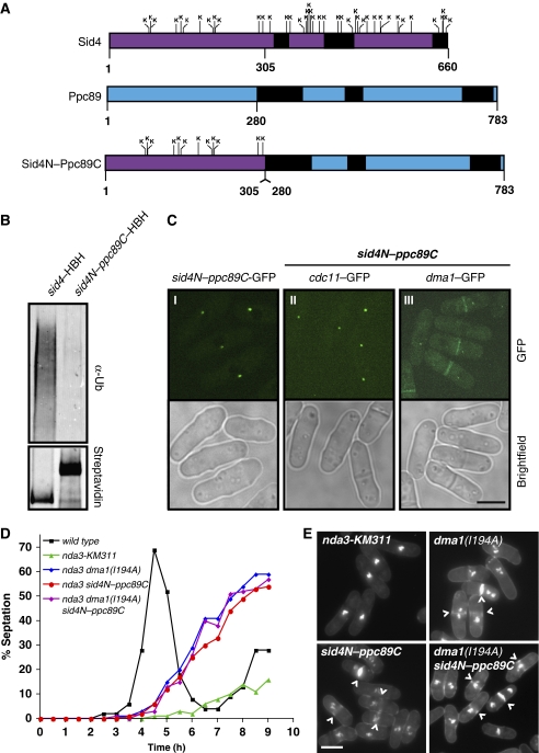
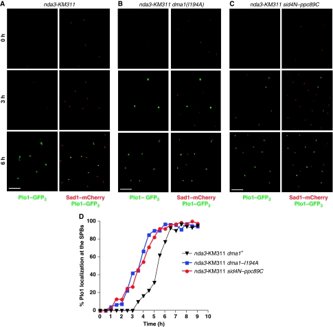
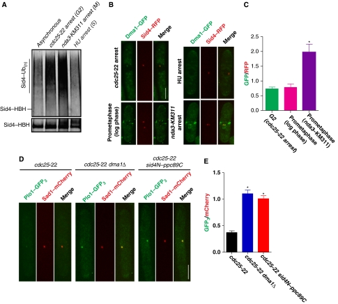
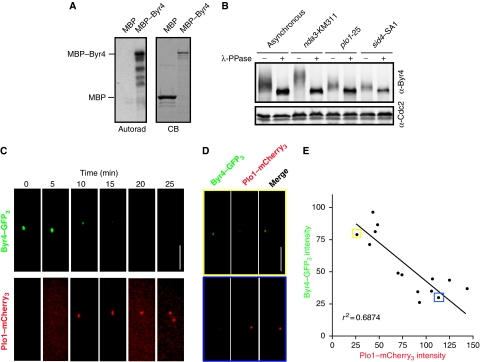
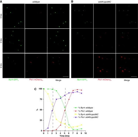
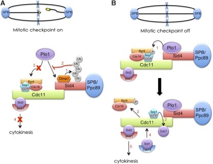
Proper cell division requires strict coordination between mitotic exit and cytokinesis. In the event of a mitotic error, cytokinesis must be inhibited to ensure equal partitioning of genetic material. In the fission yeast, Schizosaccharomyces pombe, the checkpoint protein and E3 ubiquitin ligase, Dma1, delays cytokinesis by inhibiting the septation initiation network (SIN) when chromosomes are not attached to the mitotic spindle. To elucidate the mechanism by which Dma1 inhibits the SIN, we screened all SIN components as potential Dma1 substrates and found that the SIN scaffold protein, Sid4, is ubiquitinated in vivo in a Dma1-dependent manner. To investigate the role of Sid4 ubiquitination in checkpoint function, a ubiquitination deficient sid4 allele was generated and our data indicate that Sid4 ubiquitination by Dma1 is required to prevent cytokinesis during a mitotic checkpoint arrest. Furthermore, Sid4 ubiquitination delays recruitment of the Polo-like kinase and SIN activator, Plo1, to spindle pole bodies (SPBs), while at the same time prolonging residence of the SIN inhibitor, Byr4, providing a mechanistic link between Dma1 activity and cytokinesis inhibition.


Conflict of interest statement

The authors declare that they have no conflict of interest.

Figures

Figure 1
Figure 1
The SIN scaffold, Sid4, is ubiquitinated in vivo. (A) Schematic diagram of Dma1 protein with relative positions of Dma1 FHA and RF domains and the I194A point mutation indicated. (B) In vitro ubiquitination assay using an E1-activating enzyme, the human E2-conjugating enzyme, Ubc13/Uev1a and either dma1–HA3–TAP or dma1(I194A)–HA3–TAP purified from S. pombe lysates arrested by the nda3-KM311 mutation. (C) List of SIN and SPB proteins screened for in vivo ubiquitination. (D) In vivo ubiquitination assay of proteins listed in C. Each protein was purified from checkpoint-activated cells (nda3-KM311) and visualized by immunoblot using fluorescently labelled streptavidin (bottom panels) and a Ubiquitin antibody (top panels).
Figure 2
Figure 2
Sid4 ubiquitination requires Dma1 function. (A) Schematic diagram of Dma1 domains and positions of the R64A and I194A mutations. The R64A mutation prevents interaction with phosphothreonine motifs and I196A inactivates ubiquitin ligase activity. (B) Localization of dma1–GFP (panel I), dma1(R64A)–GFP (panel II) and dma1(I194A)–GFP (panel III) in cells growing in log phase. Scale bar, 5 μm. (C) Spindle checkpoint assay. Cells of the indicated strains were synchronized at 32°C in G2 by centrifugal elutriation, shifted to 18°C, and the septation index of each strain determined every 30 min for 9 h. (D) In vivo ubiquitination status of Sid4–HBH in nda3-KM311 dma1Δ or nda3-KM311 dma1 mutants.
Figure 3
Figure 3
Sid4 ubiquitination is required to maintain a checkpoint arrest. (A) Schematic diagrams of Sid4 with relative positions of all 49 lysines (top), Ppc89 (middle) and the Sid4N–Ppc89C fusion mutant (bottom). Predicted coiled-coil regions are shown in black. (B) In vivo ubiquitination of Sid4–HBH and Sid4N–Ppc89C–HBH. (C) Localization of Sid4N–Ppc89C–GFP (panel I), Cdc11–GFP (panel II) and Dma1–GFP (panel III) in sid4N–ppc89C–HBH mutant cells. Scale bar, 5 μm. (D) Spindle checkpoint assay. Cells of the indicated strains were blocked at 32°C in S phase with hydroxyurea, released into hydroxyurea-free media at 18°C, and the septation index of each strain was determined every 30 min for 9 h. (E) Cells from each of the strains examined in 3D at the 7 h time point stained with methyl blue, which stains the septa, and DAPI, which stains DNA. (^) indicate septated cells that have bypassed the checkpoint. Scale bar, 5 μm.
Figure 4
Figure 4
Sid4 ubiquitination delays Plo1 recruitment to the SPBs when the spindle checkpoint is activated. (AC) nda3-KM311 (A), nda3-KM311 dma1(I194A) (B) or nda3-KM311 sid4N–ppc89C (C) cells were synchronized at 32°C in G2 by lactose gradient sedimentation, released to 18°C to activate the spindle checkpoint, and Plo1-GFP3 and Sad1-mCherry localization at the SPBs were imaged periodically for 9 h. In each panel, the images on the left show Plo1–GFP3 localization alone and the images on the right show merged images of Plo1–GFP3 colocalized with Sad1–mCherry at each of the times indicated. Scale bar, 10 μm. (D) The kinetics of Plo1 recruitment to SPBs was measured for each of the strains shown by calculating the percentage of cells with Plo1–GFP3 on SPBs at each time point.
Figure 5
Figure 5
Sid4 ubiquitination prevents Plo1 recruitment to SPBs during interphase. (A) In vivo ubiquitination of Sid4–HBH in asynchronous cells or cells arrested in G2 (cdc25-22), prometaphase (nda3-KM311) or S phase (hydroxyurea; HU). (B) Representative images showing Dma1–GFP and Sid4–RFP localization in a G2 arrest (cdc25-22 arrest), an S-phase arrest (HU arrest), prometaphase cell growing in log phase, and a mitotic arrest when the checkpoint is active (nda3-KM311 arrest). Scale bar, 5 μm. (C) Quantitation of relative Dma1–GFP/Sid4-RFP intensity ratios for each of the cell cycle stages shown in B plotted as arbitrary units. For each cell cycle stage, Dma1–GFP and Sid4–RFP intensities were measured for at least 20 cells and averaged; error bars represent standard error of the mean, *P<0.05. (D) Representative images showing Plo1–GFP3 and Sad1–mCherry localization at SPBs during a cdc25-22 arrest in wild type (left panels), dma1Δ (middle panels) and sid4N–ppc89C (right panels) cells. Scale bar, 5 μm. (E) Quantitation of relative Plo1–GFP3/Sad1–mCherry intensity ratios at SPBs for each of the strains shown in D plotted in arbitrary units. For each strain, Plo1–GFP3 and Sad1–mCherry intensities were measured for at least 20 cells and averaged; error bars represent standard error of the mean, *P<0.05.
Figure 6
Figure 6
Byr4 is a potential Plo1 target. (A) Left, autoradiograph of recombinant MBP and MBP–Byr4 phosphorylated in vitro by Plo1 kinase. Right, Coomassie blue (CB) gel of purified MBP and MBP–Byr4 proteins. (B) Gel shifts of endogenous Byr4 immunoprecipitated from asynchronous, nda3-KM311, plo1-25 or sid4-SA1 temperature-sensitive cells, which were synchronized in S phase by hydroxyurea and released at the restrictive temperature. Immunoprecipitates were treated with (+) or without (−) λ-phosphatase and detected by immunoblotting using an anti-Byr4 serum. (C) A Byr4–GFP3 Plo1–mCherry3 strain was imaged via time-lapse microscopy and a representative montage is depicted. (D) A Byr4–GFP3 Plo1–mCherry3 strain was grown to log phase and imaged. Top and bottom panels show representative images of cells in which Byr4–GFP3 or Plo1–mCherry3, respectively, localization to the SPB predominates. (E) Byr4–GFP3 and Plo1–mCherry3 fluorescence intensities were measured and plotted against each other. A linear regression analysis was performed to calculate the best-fit line, r2=0.687. The data points boxed in yellow and blue represent the intensity calculations for the top and bottom panels shown in D, respectively.
Figure 7
Figure 7
Sid4 ubiquitination is required to prolong Byr4 residence on SPBs when a mitotic checkpoint is activated. (A, B) nda3-KM311 (A) and nda3-KM311 sid4N–ppc89C (B) cells were synchronized in G2 by lactose gradient sedimentation, shifted to 18°C to activate the spindle checkpoint, and Byr4–GFP3 and Plo1–mCherry3 localizations at the SPBs were imaged periodically for 9 h. Representative images of Byr4–GFP3, Plo1–mCherry3 and the merged images are shown for the times indicated. Scale bar, 10 μm. (C) At each time point, the percentage of cells with Byr4–GFP3 and Plo1–mCherry3 were calculated and plotted over time. Dashed lines represent the time in which the plots for Byr4 and Plo1 intersect (∼5 h for nda3-KM311 cells and ∼2 h for nda3-KM311 sid4N–ppc89C cells).
Figure 8
Figure 8
Model of Dma1 inhibition of the SIN during a mitotic checkpoint. (A) Proposed mechanism of Dma1 inhibition of the SIN when the mitotic checkpoint is active. (B) Mechanism of SIN activation when chromosomes are properly attached to the mitotic spindle and the checkpoint is satisfied.

Similar articles

Cited by

References

    1. Alexandru G, Zachariae W, Schleiffer A, Nasmyth K (1999) Sister chromatid separation and chromosome re-duplication are regulated by different mechanisms in response to spindle damage. EMBO J 18: 2707–2721 - PMC - PubMed
    1. Bahler J, Steever AB, Wheatley S, Wang Y, Pringle JR, Gould KL, McCollum D (1998) Role of polo kinase and Mid1p in determining the site of cell division in fission yeast. J Cell Biol 143: 1603–1616 - PMC - PubMed
    1. Bardin AJ, Amon A (2001) Men and sin: what's the difference? Nat Rev Mol Cell Biol 2: 815–826 - PubMed
    1. Beltraminelli N, Murone M, Simanis V (1999) The S. pombe zfs1 gene is required to prevent septation if mitotic progression is inhibited. J Cell Sci 112(Pt 18): 3103–3114 - PubMed
    1. Bothos J, Summers MK, Venere M, Scolnick DM, Halazonetis TD (2003) The Chfr mitotic checkpoint protein functions with Ubc13-Mms2 to form Lys63-linked polyubiquitin chains. Oncogene 22: 7101–7107 - PubMed

Publication types

MeSH terms

Substances