. 2011 Jan 20;469(7330):402-6.
doi: 10.1038/nature09600. Epub 2010 Dec 5.

Development of asymmetric inhibition underlying direction selectivity in the retina

Affiliations

Development of asymmetric inhibition underlying direction selectivity in the retina

Wei Wei et al. Nature. .

Abstract

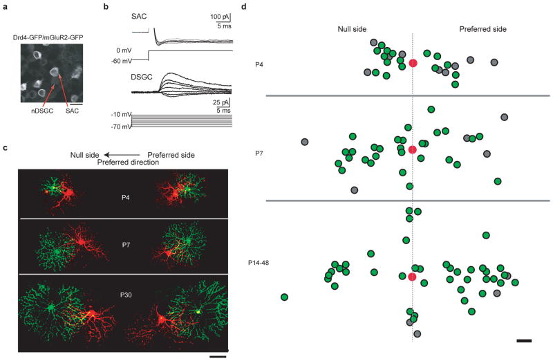
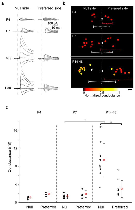
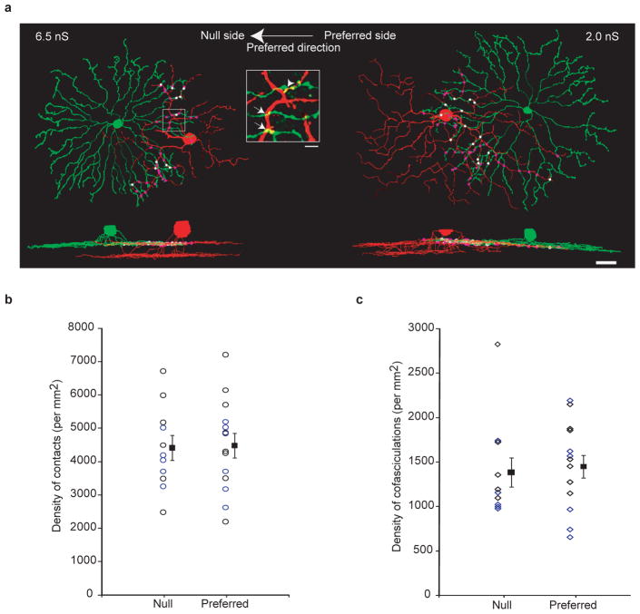
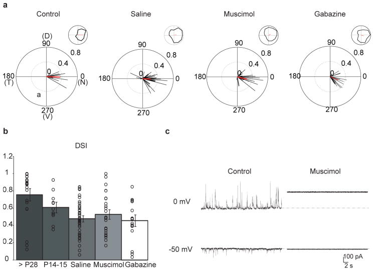
Establishing precise synaptic connections is crucial to the development of functional neural circuits. The direction-selective circuit in the retina relies upon highly selective wiring of inhibitory inputs from starburst amacrine cells (SACs) onto four subtypes of ON-OFF direction-selective ganglion cells (DSGCs), each preferring motion in one of four cardinal directions. It has been reported in rabbit that the SACs on the 'null' sides of DSGCs form functional GABA (γ-aminobutyric acid)-mediated synapses, whereas those on the preferred sides do not. However, it is not known how the asymmetric wiring between SACs and DSGCs is established during development. Here we report that in transgenic mice with cell-type-specific labelling, the synaptic connections from SACs to DSGCs were of equal strength during the first postnatal week, regardless of whether the SAC was located on the preferred or null side of the DSGC. However, by the end of the second postnatal week, the strength of the synapses made from SACs on the null side of a DSGC significantly increased whereas those made from SACs located on the preferred side remained constant. Blocking retinal activity by intraocular injections of muscimol or gabazine during this period did not alter the development of direction selectivity. Hence, the asymmetric inhibition between the SACs and DSGCs is achieved by a developmental program that specifically strengthens the GABA-mediated inputs from SACs located on the null side, in a manner not dependent on neural activity.


Conflict of interest statement

The authors declare no competing financial interests.

Figures

Figure 1
Figure 1. nDSGCs receive direct GABAergic inputs from SACs located on the null and the preferred side from P4 until adult
a, Fluorescence image of the ganglion cell layer from a P30 Drd4–GFP/mGluR2–GFP mouse, showing the bright membrane-bound GFP expressed under the mGluR2 promoter in the SACs and the dim cytoplasmic GFP driven by the Drd4 promoter in the nDSGC. Scale bar, 25 μm. b, Paired whole-cell voltage-clamp recordings of GABAergic currents in a P4 nDSGC (lower traces) evoked by depolarization of a SAC from the null side (upper traces) in the presence of the NMDA (N-methyl-D-aspartate) receptor antagonist D(−)-2-amino-5-phosphonovaleric acid (AP5), α-amino-3-hydroxy-5-methyl-4-isoxazole propionic acid (AMPA)/kainate receptor antagonist 6,7-dinitroquinoxaline-2,3-dione (DNQX) and α4-containing nicotinic acetylcholine receptor antagonist dihydro-β-erythroidine (DHβE). SACs were depolarized from −60 to 0 mV, which reliably evoked an inward current in SACs. The postsynaptic GABAergic currents were recorded in DSGCs at different holding potentials to determine the current–voltage relationship of the conductance. c, Example images of synaptically connected, dye-filled SAC–DSGC pairs at P4, P7 and P30. The left-hand side shows pairs with SACs (green) located on the null side of the DSGCs (red). The right-hand side shows preferred-side pairs. Scale bar, 50 μm. d, Soma locations of the GABAergically connected SAC–nDSGC pairs along the null-preferred axis during development. Red spots represent the positions of DSGC cell bodies. The positions of SAC cell bodies that form GABAergic synapses with their respective nDSGCs are shown as green spots; the SAC cell bodies that were not connected to nDSGCs are shown as grey spots. All pairs had overlapping dendritic fields. Scale bar, 25 μm
Figure 2
Figure 2. GABAergic conductance in the null-side SAC–nDSGC pairs strengthens during the second postnatal week
a, Postsynaptic GABAergic currents in nDSGCs recorded at holding potentials between −70 and −10 mV in response to depolarization (as in Fig. 1b) of null-side (left) and preferred-side (right) SACs at P4, P7, P14 and P30. b, Relative soma positions of SAC–nDSGC pairs used for conductance analysis at P4, P7 and P14–48. Open circles represent nDSGC cell bodies. Filled circles are SAC somas colour-coded for conductance strength normalized to the maximum value across all ages. Dashed lines illustrate average dendritic arborization diameter, centred on the asterisks, for nDSGCs (white) and SACs (red; asterisks represent average soma locations). Scale bar, 25 μm. c, Summary plot of GABAergic conductances of the null- and preferred-side SAC–nDSGC pairs at P4, P7 and P14–48. Individual pairs and mean ± s.d. are shown. One-way analysis of variance: P < 0.0001; t-test: *P < 0.0001, **P = 0.0003.
Figure 3
Figure 3. Dendritic contacts and cofasciculations between SACs and nDSGCs occur at similar densities for the null- and preferred-side pairs
a, NEUROLUCIDA reconstructions of the dendrites from the on sublamina and side views of the complete dendritic arborizations from a null-side (left) and a preferred-side (right) pair of SACs and nDSGCs. Dots represent dendritic contacts, with cofasciculation segments coloured white and the rest coloured purple. The GABAergic conductances for the null- and preferred-side pairs are indicated. Scale bar, 25 μm. Inset, fluorescence image of the outlined region showing crossing contacts (arrows) and cofasciculation (arrowhead). Scale bar, 5 μm. b, Summary plot of the density of total contacts between DSGCs and distal SAC processes (roughly the outer third) from the null or preferred side from P14 to P48. Individual pairs and mean ± s.d. are shown. The data points for P28 and later are coloured blue, and the ones for before P28 are coloured black. c, Summary plot of the density of cofasciculations between nDSGCs and distal SAC processes from the same pairs as in b. Null- and preferred-side groups are not significantly different in b and c. P > 0.7, t-test.
Figure 4
Figure 4. Intraocular injections of muscimol or gabazine do not alter direction selectivity in nDSGCs
a, The normalized spike vector sums of nDSGCs in response to drifting gratings of 12 directions from P14–15 Drd4–GFP mice that received either no treatment (control) or intraocular injections of saline, muscimol or gabazine from P6 to P12. D, dorsal; N, nasal; T, temporal; V, ventral. The red lines are mean vector sums of all cells in each group. Insets, examples of normalized tuning curves of single cells, with corresponding vector sums represented as red lines of nDSGCs from each group. Control: n = 4 mice, 12 cells; saline: n = 11 mice, 43 cells; muscimol: n = 12 mice, 25 cells; gabazine: n = 4 mice, 17 cells. b, Summary plot of direction selectivity index (DSI) for adult (>P28), P14–15 untreated, saline, muscimol and gabazine-treated groups. Bars show mean ± s.e.; open circles represent individual cells. Adult data are reproduced from ref. . c, Example traces from whole-cell voltage-clamp recordings of inhibitory (upper traces, VH = 0 mV) and excitatory (lower traces, VH = −50 mV) currents from a P14 nDSGC in drug-free artificial cerebrospinal fluid (control, left) or artificial cerebrospinal fluid containing 100 μM muscimol (right). Deflections from baseline correspond to spontaneous synaptic currents. At depolarized potentials, application of muscimol activated a tonic current, which was measured as a change in the baseline holding current.

Comment in

Similar articles

Cited by

References

    1. Euler T, Detwiler PB, Denk W. Directionally selective calcium signals in dendrites of starburst amacrine cells. Nature. 2002;418:845–852. - PubMed
    1. Demb JB. Cellular mechanisms for direction selectivity in the retina. Neuron. 2007;55:179–186. - PubMed
    1. Fried SI, Munch TA, Werblin FS. Mechanisms and circuitry underlying directional selectivity in the retina. Nature. 2002;420:411–414. - PubMed
    1. Barlow HB, Levick WR. The mechanism of directionally selective units in rabbit’s retina. J Physiol (Lond) 1965;178:477–504. - PMC - PubMed
    1. Oyster CW. The analysis of image motion by the rabbit retina. J Physiol (Lond) 1968;199:613–635. - PMC - PubMed

Publication types

MeSH terms