. 2011 Jan;13(1):87-94.
doi: 10.1038/ncb2139. Epub 2010 Dec 5.

CDK1-dependent phosphorylation of EZH2 suppresses methylation of H3K27 and promotes osteogenic differentiation of human mesenchymal stem cells

Affiliations

CDK1-dependent phosphorylation of EZH2 suppresses methylation of H3K27 and promotes osteogenic differentiation of human mesenchymal stem cells

Yongkun Wei et al. Nat Cell Biol. 2011 Jan.

Abstract

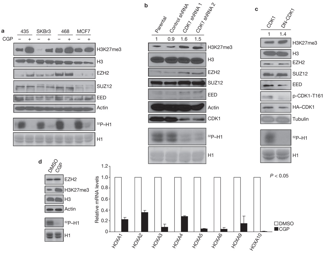
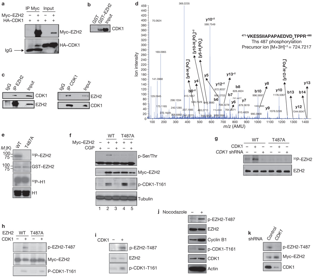
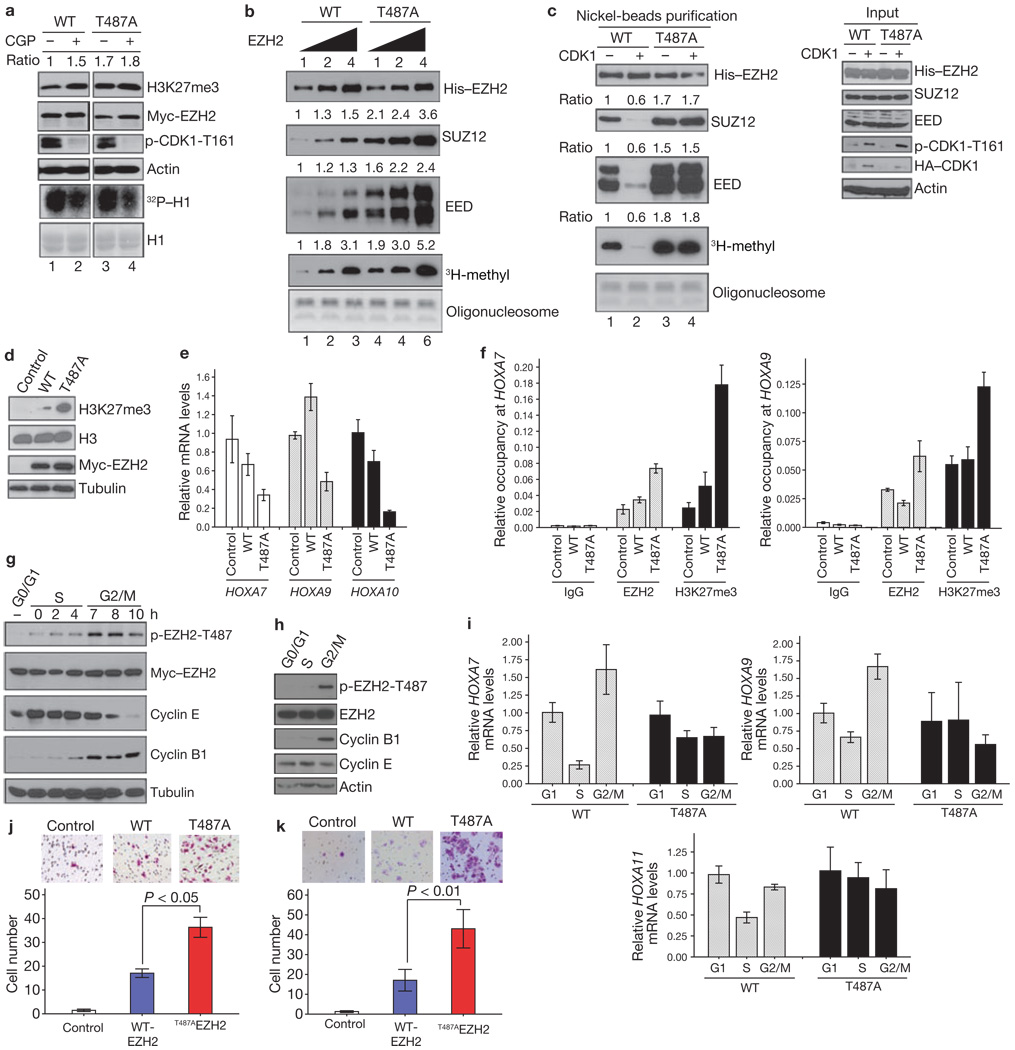
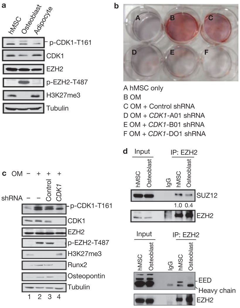
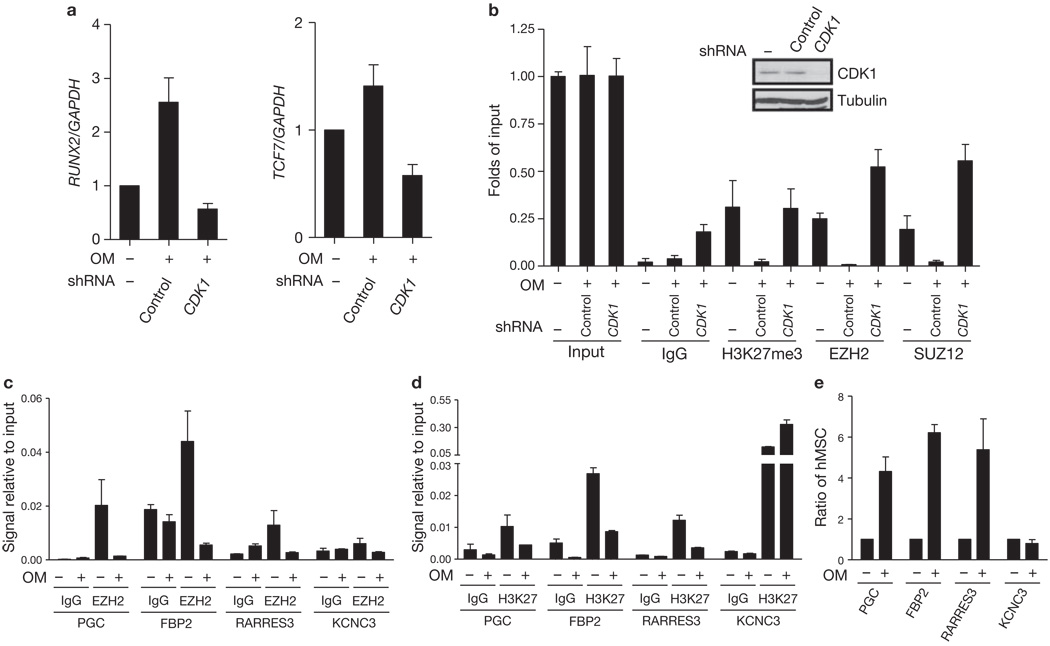
Enhancer of zeste homologue 2 (EZH2) is the catalytic subunit of Polycomb repressive complex 2 (PRC2) and catalyses the trimethylation of histone H3 on Lys 27 (H3K27), which represses gene transcription. EZH2 enhances cancer-cell invasiveness and regulates stem cell differentiation. Here, we demonstrate that EZH2 can be phosphorylated at Thr 487 through activation of cyclin-dependent kinase 1 (CDK1). The phosphorylation of EZH2 at Thr 487 disrupted EZH2 binding with the other PRC2 components SUZ12 and EED, and thereby inhibited EZH2 methyltransferase activity, resulting in inhibition of cancer-cell invasion. In human mesenchymal stem cells, activation of CDK1 promoted mesenchymal stem cell differentiation into osteoblasts through phosphorylation of EZH2 at Thr 487. These findings define a signalling link between CDK1 and EZH2 that may have an important role in diverse biological processes, including cancer-cell invasion and osteogenic differentiation of mesenchymal stem cells.


Figures

Figure 1
Figure 1
CDK1 negatively regulates H3K27 trimethylation. (a) Top: 435, SKBr3, 468 and MCF7 cells were treated with CGP74514A as indicated, and the lysates were analysed by immunoblot using antibodies against the specified proteins. Bottom: in vitro kinase assay. CDK1, immunoprecipitated from the cell lines treated with CGP74514A as indicated at the top, was incubated with H1 and [γ-32P]ATP. Reaction products were resolved by SDS–PAGE and visualized by autoradiography (equal loading of H1 was assessed by Coomassie-stained gel shown at the bottom). (b) Lysates from MCF7 cells infected with lentiviruses expressing control or two different CDK1 shRNA were immunoblotted with antibodies against the indicated proteins. Relative intensities of the H3K27me3 bands are shown, normalized to the H3K27me3 band from parental MCF7 cells. Bottom: in vitro kinase assay, performed as in a, with CDK1 immunoprecipitated from cells treated as indicated at the top. (c) Lysates of 293T cells transfected with plasmids encoding cyclin B, and CDK1 or dominant-negative mutant CDK1 (DN-CDK1) were immunoblotted with antibodies against the indicated proteins. Relative intensities of the H3K27me3 bands are shown, normalized to the H3K27me3 band from 293T cells transfected with plasmid encoding CDK1. p-CDK1-T161; CDK1 phosphorylated at Thr 161. Bottom: in vitro kinase assay, performed as in a, with CDK1 immunoprecipitated from cells transfected as indicated at the top. (d) Left top: immunoblot of lysate from HEK293 cells treated with DMSO or CGP using antibodies against the indicated proteins. Left bottom: in vitro kinase assay, performed as in a, with CDK1 immunoprecipitated from cells treated as indicated at the top. Right: analysis of mRNA levels of HOXA families by qRT–PCR after treatment of HEK293 cells with DMSO or CGP74514A. Data are means ± s.e.m. (n = 3). Uncropped images of blots are shown in Supplementary Information, Fig. S6.
Figure 2
Figure 2
CDK1 interacts with, and phosphorylates, EZH2 at Thr 487. (a) Lysates from 293T cells, transfected with plasmids encoding Myc–EZH2 and HA–CDK1 as indicated, were immunoprecipitated (IP) using anti-Myc and analysed by immunoblot using anti-Myc or anti-HA. IgG; immunoglobulin G. (b) Pulldowns. GST and GST–EZH2 were incubated with lysate from HeLa cells. Bound CDK1 was detected by immunoblotting. (c) Lysate from MCF7 cells was immunoprecipitated with antibodies against EZH2 (left) or CDK1 (right), and analysed by immunoblotting. (d) Cyclin B, CDK1 and GST–EZH2 were subjected to an in vitro kinase assay and analysed by mass spectrometry. The spectrum of the charged ion (m/z 724.7217) shows that Thr 487 is phosphorylated (lower case p) in the indicated peptide (top right). b ions, fragmentation ions containing the amino terminus of the peptide; y ions, fragmentation ions containing the carboxy terminus of the peptide. (e) In vitro kinase assay with CDK1, cyclin B, and wild-type GST–EZH2 (WT) or GST–EZH2T487A. Phosphorylation of EZH2 and H1 was visualized by autoradiography, and loading of GST–EZH2 and H1 was assessed by Coomassie-stained gel. (f) 293T cells were transfected with plasmids encoding wild-type Myc–EZH2 or Myc–EZH2T487A and treated with CDK1 inhibitor CGP74514A or DMSO. Lysates were immunoprecipitated with anti-Myc and analysed by immunoblotting. (g) MCF7 cells stably expressing wild-type Myc–EZH2 or Myc–EZH2T487A were transfected with control vector or plasmids encoding CDK1 and cyclin B, and infected with lentivirus expressing CDK1 shRNA, as indicated. Cells were labelled with [32P]-orthophosphate, EZH2 was immunoprecipitated from lysates with anti-Myc and analysed by autoradiography. Immunoblotting was used to confirm equal loading of EZH2 (bottom). (h) HeLa cells expressing Myc–EZH2 or Myc–EZH2T487A were transfected with plasmid encoding CDK1 and cyclin B, or control vector. Cell lysates were immunoprecipitated with anti-Myc and immunoblotted. (i) Lysates of HeLa cells transfected with plasmid encoding CDK1 and cyclin B, or control vector, were immunoprecipitated with anti-EZH2, and immunoblotted. (j) Immunoblot of lysates from HeLa cells treated with Nocodazole as indicated. Cell lysates were immunoprecipitated with anti-EZH2. (k) MCF7 cells stably expressing wild-type Myc–EZH2 were infected with lentivirus expressing control or CDK1 shRNA. Cell lysates were immunoprecipitated with anti-Myc and immunoblotted. Uncropped images of blots are shown in Supplementary Information, Fig. S6.
Figure 3
Figure 3
CDK1-mediated phosphorylation of EZH2 promotes disassociation of EZH2 from SUZ12 and EED and suppresses EZH2 HMTase activity. (a) HeLa cells were transfected with plasmids encoding wild-type EZH2 or EZH2T487A and treated with CGP74514A as indicated. Top: immunoblot. Relative intensity of the H3K27me3 bands is indicated. Bottom: Autoradiograph and immunoblot of in vitro kinase assay using H1 as substrate. (b) In vitro histone methyltransferase assay. PRC2 complexes were purified from MCF7 cell lines stably expressing wild-type Myc-His–EZH2 or Myc-His–EZH2T487A and 0.5, 1 and 2 µg were used in the assay. Top: immunoblots of purified proteins. Bottom: proteins were incubated with oligonucleosome and 3H-labelled S-adenosylmethionine. Methylation was assessed by autoradiography and oligonucleosome loading by Coomassie-stained gel. Relative intensities of bands are indicated at the top of each panel. (c) MCF7 cell lines as in b were transfected with control vector or plasmids encoding HA–CDK1. Left: immunoblots of experiment performed as in b. Right: immunoblot of lysates from MCF7 cells used in protein purification. (d) Immunoblot of lysates from stable MCF7 transfectants, established by transfection of cells with control vector or plasmids encoding Myc–EZH2 or Myc–EZH2T487A. (e) qRT–PCR of HOXA genes in MCF7 cell lines described in d. Data are means ± s.d. from three individual experiments. (f) Quantitative chromatin immunoprecipitation analysis on HOXA7 and HOXA9 promoters in MCF7 cell lines as described in d. Data are means ± s.d. from three individual experiments. (g) Immunoblot of MCF7 cell lines stably expressing wild-type EZH2, treated by serum starvation (to collect cells at G0/G1 phase) or by double thymidine blockage and release (to collect cells at S and G2/M phases). (h) Immunoblot of HeLa cells treated as in g. (i) qRT–PCR of HOXA gene expression in MCF7 stable cell lines treated as in g. Data are means ± s.d. from three individual experiments. (j) Images and quantification of cell migration of MCF7 stable cell lines. Data are means ± s.e.m. from three individual experiments. (k) Images and quantification of cell invasion of MCF7 stable cell lines. Data are means ± s.e.m. from three individual experiments. Uncropped images of blots are shown in Supplementary Information, Fig. S6.
Figure 4
Figure 4
Phosphorylation of EZH2 by CDK1 promotes osteogenic differentiation of human mesenchymal stem cells. (a) Osteoblast differentiation medium induces activation of CDK1 in hMSCs. Immunoblot of lysates from undifferentiated cells or cells differentiated into osteoblasts or adipocytes. (b) hMSCs were left untreated or were treated with osteoblast differentiation medium (OM) and shRNA as indicated. Alizarin Red S staining was performed at day 7. Differentiated stem cells positive for Alizarin Red S are stained red. (c) Effect of CDK1 knockdown on the expression of osteogenic-specific genes in hMSCs. Cells were cultured in control medium or osteoblast differentiation medium, and infected with lentiviruses expressing control or CDK1 shRNA as indicated. Cell lysates were subjected to immunoblot analysis. (d) Disruption of PRC2 complex after osteogenic differentiation. Lysates from cells undifferentiated or differentiated into osteoblasts were immunoprecipitated with EZH2 antibody and subjected to immunoblot analysis as indicated.
Figure 5
Figure 5
CDK1 regulates EZH2-target gene expression in human mesenchymal stem cells. (a) Effect of CDK1 knockdown on the expression of EZH2-target genes in hMSCs. Cells were cultured in control medium or osteoblast differentiation medium with or without CDK1 shRNA. mRNA levels of Runx2 (left) and TCF7 (right) were measured by qRT–PCR, and are calculated relative to GAPDH expression. Data are means ± s.e.m. (n = 3). (b) Effect of CDK1 knockdown on the binding of EZH2 to Runx2 gene promoter in hMSCs. Cells were cultured in control medium or osteoblast differentiation medium with or without CDK1 shRNA infection. Quantitative chromatin immunoprecipitation was performed using antibodies against the indicated proteins on the Runx2 promoter. Levels of CDK1 are shown by immunoblot (top). (c, d) Four genes shown to have differential EZH2 binding after osteogenic differentiation from a genome-wide ChIP-on-chip assay were randomly selected for ChIP assay using antibodies against EZH2 (c) and H3K27me3 (d). hMSCs were cultured in control medium or osteoblast differentiation medium. (e) The expression of the four genes analysed in c and d was assessed by qRT–PCR from hMSCs cultured in control medium or osteoblast differentiation medium.

Similar articles

Cited by

References

    1. Cao R, et al. Role of histone H3 lysine 27 methylation in Polycomb-group silencing. Science. 2002;298:1039–1043. - PubMed
    1. Kuzmichev A, Nishioka K, Erdjument-Bromage H, Tempst P, Reinberg D. Histone methyltransferase activity associated with a human multiprotein complex containing the Enhancer of Zeste protein. Genes Dev. 2002;16:2893–2905. - PMC - PubMed
    1. Cao R, Zhang Y. The functions of E(Z)/EZH2-mediated methylation of lysine 27 in histone H3. Curr. Opin. Genet. Dev. 2004;14:155–164. - PubMed
    1. Varambally S, et al. The polycomb group protein EZH2 is involved in progression of prostate cancer. Nature. 2002;419:624–629. - PubMed
    1. Kleer CG, et al. EZH2 is a marker of aggressive breast cancer and promotes neoplastic transformation of breast epithelial cells. Proc. Natl Acad. Sci. USA. 2003;100:11606–11611. - PMC - PubMed

Publication types

MeSH terms