. 2011 Jan;7(1):51-7.
doi: 10.1038/nchembio.494. Epub 2010 Dec 5.

Structural basis for regulation of the Crk signaling protein by a proline switch

Affiliations

Structural basis for regulation of the Crk signaling protein by a proline switch

Paramita Sarkar et al. Nat Chem Biol. 2011 Jan.

Abstract

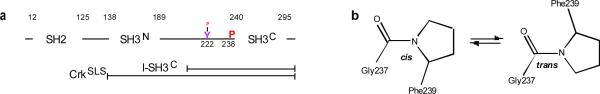
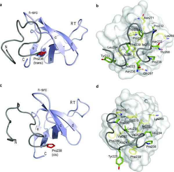
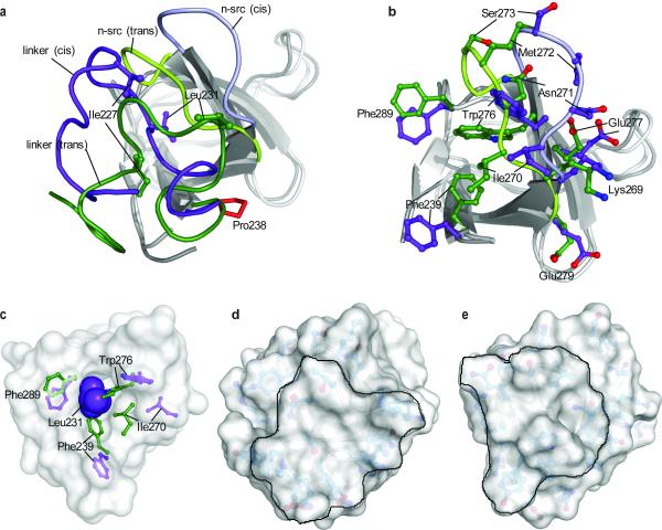
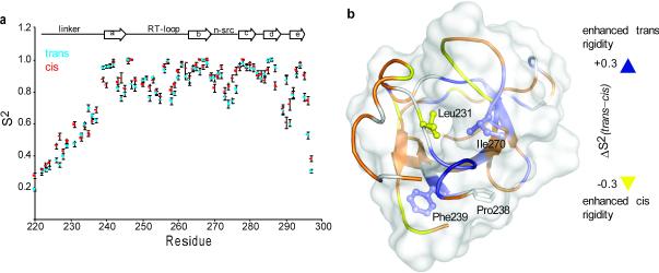
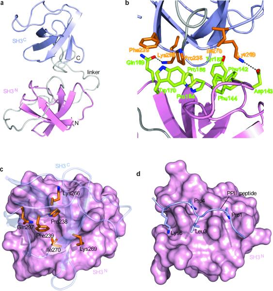
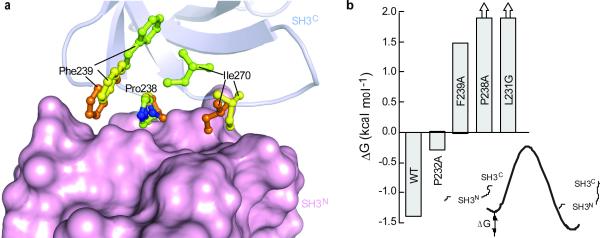
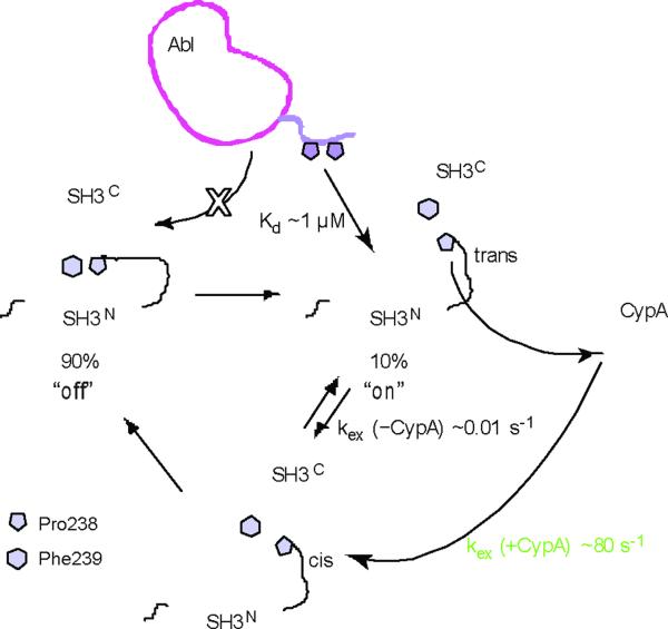
Proline switches, controlled by cis-trans isomerization, have emerged as a particularly effective regulatory mechanism in a wide range of biological processes. Here we report the structures of both the cis and trans conformers of a proline switch in the Crk signaling protein. Proline isomerization toggles Crk between two conformations: an autoinhibitory conformation, stabilized by the intramolecular association of two tandem SH3 domains in the cis form, and an uninhibited, activated conformation promoted by the trans form. In addition to acting as a structural switch, the heterogeneous proline recruits cyclophilin A, which accelerates the interconversion rate between the isomers, thereby regulating the kinetics of Crk activation. The data provide atomic insight into the mechanisms that underpin the functionality of this binary switch and elucidate its remarkable efficiency. The results also reveal new SH3 binding surfaces, highlighting the binding versatility and expanding the noncanonical ligand repertoire of this important signaling domain.


Figures

Figure 1
Figure 1. Domain organization of Crk
(a) Schematic diagram of the domain organization of Crk. Pro238, which undergoes cis–trans isomerization, and the tyrosine residue (Tyr222) that becomes phosphorylated by Abl are indicated. (b) Schematic of cis–trans isomerization about the prolyl Gly237–Pro238 bond.
Figure 2
Figure 2. Structural characterization of the trans and cis Crk l-SH3C conformers
(a,b) Structure of the Crk l-SH3C polypeptide in the trans conformation and (c,d) in the cis conformation. In (a and c) the SH3C domain is colored light blue and the linker is colored grey. Pro238 is shown in red. N and C denote the N- and C-terminus. In (b and d) the SH3C domain is displayed as a semi-transparent surface and residues involved in the linker–SH3C interactions are highlighted. Linker and SH3C residues are colored green and yellow, respectively. Dotted lines denote hydrogen bonding.
Figure 3
Figure 3. Structural comparison of the trans and cis isomers
(a) Superposition of the trans and cis conformers of l-SH3C on the SH3C domain (residues 239–295). The SH3C domain in the trans and cis conformation is in light and dark grey, respectively. The linker adopts a very different structure in the trans (green) and the cis (purple) conformation. Representative residues at the linker are indicated. The conformation of the n-src loop is distinct in the trans (green) and the cis (purple) conformer. (b) Effect of cis–trans isomerization on the structure of SH3C. The side-chain conformation of select residues is shown. The side chain of Phe239 is solvent exposed in the cis isomer (purple) whereas it is buried in the trans isomer (green). Superposition of the structures and color code are as in (a). (c) Leu231 is completely buried into a hydrophobic pocket in SH3C in the cis conformation. The side-chain conformation of residues lining the hydrophobic pocket is shown in the cis (purple) and trans (green) isomers.(d,e) Solvent-accessible surface presentation of the trans (d) and cis (e) conformations. The models are rotated ~60° about the z axis relative to the models in (a) with the linker facing the reader. The surface presented by the linker is delineated.
Figure 4
Figure 4. Dynamic characterization of the trans and cis isomers
(a) N-H bond order parameters, S2, plotted as a function of the primary sequence, for the trans (blue) and cis (red) conformers of l-SH3C. (b) Changes in order parameters, ΔS2, between the trans and the cis conformers mapped using a gradient color code on the structure of cis l-SH3C. ΔS2 is given as S2 (trans) – S2 (cis), so positive ΔS2 values denote enhanced rigidity of the protein backbone in the trans over the cis conformation.
Figure 5
Figure 5. Structural characterization of CrkSLS in the closed, autoinhibited conformation
(a) Lowest-energy structure of CrkSLS shown as cartoon with SH3N in pink, SH3C in light blue, and linker in grey. (b) Close-up view of the SH3N–SH3C interface. SH3N and SH3C residues are colored green and orange, respectively. (c) View from above SH3C, which is shown as transparent blue cartoon. SH3N is displayed as pink solvent-accessible surface. SH3C residues interacting with SH3N are shown as orange sticks. (d) Structure of SH3N in complex with a consensus PPII peptide (P-x-P-L-x-K). The orientation of SH3N in (c) and (d) is the same. Comparison of (c) and (d) show that the PPII-binding site in CrkSLS is not accessible.
Figure 6
Figure 6. Structural basis for conformer-specific SH3N–SH3C interaction in CrkSLS
(a) Superposition of the cis and trans l-SH3C and CrkSLS on the SH3C domain. The side chains of Pro238, Phe239, and Ile270, residues primarily mediating the SH3N–SH3C interaction, are shown in green for trans and yellow for cis l-SH3C and orange for CrkSLS. SH3N is shown as solvent-accessible surface. (b) Effect of single amino acid substitution on the stability of the closed conformation of CrkSLS as assessed by measuring the population of the closed and open conformations of CrkSLS by NMR. ΔG for P238A and L231G is a lower-bound limit since populations less than ~5% are beyond the NMR detection limit.
Figure 7
Figure 7. Mechanistic basis for the the regulation of Crk activity
Crk adopts predominantly (~90%) the closed, auto-inhibited conformation but a minor population (~10%) adopts the open, uninhibited conformation wherein the PPII-binding site on SH3N is accessible for binding. The P-x-L-P-x-K motif of Abl binds to SH3N with a relatively strong affinity (Kd ~1 μM). The open conformation exists as an equilibrium between the cis and the trans isomer, but only the cis one forms the closed conformation. The rates of the cis–trans interconversion are regulated by the action of CypA, which accelerates the interconversion by four orders of magnitude.

Comment in

Similar articles

Cited by

References

    1. Birge RB, Kalodimos C, Inagaki F, Tanaka S. Crk and CrkL adaptor proteins: networks for physiological and pathological signaling. Cell Commun Signal. 2009;7:13. - PMC - PubMed
    1. Feller SM. Crk family adaptors-signalling complex formation and biological roles. Oncogene. 2001;20:6348–71. - PubMed
    1. Miller CT, et al. Increased C-CRK proto-oncogene expression is associated with an aggressive phenotype in lung adenocarcinomas. Oncogene. 2003;22:7950–7. - PubMed
    1. Nishihara H, et al. Molecular and immunohistochemical analysis of signaling adaptor protein Crk in human cancers. Cancer Lett. 2002;180:55–61. - PubMed
    1. Linghu H, et al. Involvement of adaptor protein Crk in malignant feature of human ovarian cancer cell line MCAS. Oncogene. 2006;25:3547–3556. - PubMed

Publication types

MeSH terms

LinkOut - more resources