. 2011 Jan;43(1):79-84.
doi: 10.1038/ng.727. Epub 2010 Dec 5.

The coiled-coil domain containing protein CCDC40 is essential for motile cilia function and left-right axis formation

Affiliations

The coiled-coil domain containing protein CCDC40 is essential for motile cilia function and left-right axis formation

Anita Becker-Heck et al. Nat Genet. 2011 Jan.

Abstract

Primary ciliary dyskinesia (PCD) is a genetically heterogeneous autosomal recessive disorder characterized by recurrent infections of the respiratory tract associated with the abnormal function of motile cilia. Approximately half of individuals with PCD also have alterations in the left-right organization of their internal organ positioning, including situs inversus and situs ambiguous (Kartagener's syndrome). Here, we identify an uncharacterized coiled-coil domain containing a protein, CCDC40, essential for correct left-right patterning in mouse, zebrafish and human. In mouse and zebrafish, Ccdc40 is expressed in tissues that contain motile cilia, and mutations in Ccdc40 result in cilia with reduced ranges of motility. We further show that CCDC40 mutations in humans result in a variant of PCD characterized by misplacement of the central pair of microtubules and defective assembly of inner dynein arms and dynein regulatory complexes. CCDC40 localizes to motile cilia and the apical cytoplasm and is required for axonemal recruitment of CCDC39, disruption of which underlies a similar variant of PCD.


Figures

Figure 1
Figure 1. Mutation of the uncharacterized Ccdc40 gene in lnks mutant mouse embryos results in laterality defects
a–d. Heart, lung and stomach (S) from E13.5 (a,b) and E12.5 (c,d) wildtype (a,c) embryos exhibiting normal situs (NS) and lnks mutant viscera exhibiting left isomerism (b, LI), or situs inversus (d, SI). Right ventricle (RV). Heart is outlined in black, left and right lung lobes in blue and red, respectively. e. Genetic map of lnks interval on mouse chromosome 11. The number of recombination events over the number of opportunities for recombination is indicated for each polymorphic marker. D11ski16 never separated from the lnks phenotype. Within this interval are four transcription units: TBC1 domain family, member 16 (Tbc1d16), coiled-coil domain containing 40 (Ccdc40), glucosidase, alpha, acid (Gaa) and eukaryotic translation initiation factor 4A3 (Eif4a3) f–i. Ccdc40 expression in wildtype E8.0 (f), E8.25 (g), E8.5 (h) and E9.5 (i) embryos as detected by RNA in situ hybridization. Strong staining is detected in the node (arrows in f–h). j. The lnks ENU-induced mutation results in a C to A transversion (green arrow) at position 2585 in the Ccdc40 coding sequence introducing a nonsense mutation changing Valine862 to a stop codon. Mouse Ccdc40 is 63% identical and 78% similar to human CCDC40. k. The lnks mutation truncates the Ccdc40 protein within the coiled-coil domain (red). Green line in panel k indicates the peptide used to generate the anti-Ccdc40 antibody.
Figure 2
Figure 2. Loss of zebrafish ccdc40 in lok mutants or ccdc40 morpholino injected embryos produces laterality defects
a–c. Expression of ccdc40 transcript in wildtype zebrafish embryos at 75% epiboly (a) and 6 somites (b,c). Staining is detected in the dorsal forerunner cells (arrow in a), pronephric tubules (arrows in b) and otic vesicles (arrows in c). d. The predicted domain structure of the 941 amino acid zebrafish Ccdc40 protein. The lok mutation introduces a stop codon at position 778 producing a protein with a truncated C-terminal domain. Zebrafish ccdc40 is 39% and 36% identical and 60% and 58% similar to the human and mouse genes, respectively. e–h. Phenotypes of lok mutant and ccdc40MO injected embryos at 3dpf. lok mutant embryo (f) display the curly-tail phenotype compared to an unaffected sibling (e). Embryo injected with ccdc40MO1 also displays the curly-tail phenotype (g) which can be rescued by co-injection of ccdc40 mRNA (h). Insets in g and h indicate the fluorescein labeled MO was injected into both embryos. i. Quantification of left-right organ patterning in lok mutant and ccdc40MO injected embryos. SS=situs solitus; SI=situs inversus, HTX-heterotaxia, any organ pattern that is not SS or SI. j. Rescue of MO phenotypes by co-injection of ccdc40 mRNA. Heart looping was scored as an indication of left-right patterning. RLoop = rightward looping of the heart, NLoop = no heart looping (midline) and LLoop = leftward looping of the heart. Curly tail down (CTD) indicates a strong phenotype such as that pictured in g. +/++ indicates tails that were slightly kinked or bent. WT indicates indistinguishable from uninjected embryos (compare tail in h to that in e).
Figure 3
Figure 3. Loss of Ccdc40 results in ciliary defects
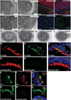
a–b. SEM showing morphology of node cilia in E8.0 wildtype (a,e) and lnks mutant (b,f) mouse embryos. Panels e and f are higher magnification views of a and b, scale bars are indicated. c,d,gh. Cilia imaging in zebrafish pronephric tubules (c,d) and Kuppfer’s vesicle (g,h). Loss of Ccdc40 function in mouse (b,f) and zebrafish embryos (d,h) results in significantly shorter cilia relative to controls. In Kupffer’s vesicle, cilia length in uninjected controls averaged 5.2µm (SD 1.486; n=589 cilia) while cilia in MO embryos were consistently shorter, averaging 3.6µm (SD 1.20; n=511 cilia; p=6×10−73 by one-tailed student t-test). Shorter cilia are also reported in lok mutants i–l. TEM analysis of cilia in the pronephros of lok mutant embyos demonstrates defects in central pair positioning (j,k) or number (l) compared to control (i). Note that outer dynein arms are not affected. m–x. Immunofluorescence analysis showing localization of endogenous Ccdc40 protein (n,q,t,w) and acetylated tubulin (m,p,s,v) in the node of E8.0 wildtype (m–o) and lnks mutant (p–r) embryos, and P21 wildtype trachea (s–u) and lnks mutant trachea (v–x) (overlay including visualization of nuclei (Hoechst staining) in o, r, u, and x.). Arrow in o points to a node cilium that was not recognized by the anti-Ccdc40 antibody. Note that Ccdc40 is not readily detectable in the 9+0 node cilium, but is present in the axonemes of multiciliated tracheal cells. Motility of node cilia was not evaluated.
Figure 4
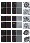
Figure 4. Localization of DNAH5, GAS11 and DNALI1 in respiratory epithelial cells from PCD patients carrying CCDC40 mutations
Immunofluorescence analyses of human respiratory epithelial cells using specific antibodies directed against the outer dynein arm heavy chain DNAH5 (a), the dynein regulating complex component GAS11 (b) and the inner dynein arm component DNALI1 (c). As control, axoneme-specific antibodies against acetylated α-tubulin (a) or α/β-tubulin (b,c) were used. Nuclei were stained with Hoechst 33342 (blue). (a) In respiratory epithelial cells from healthy probands, DNAH5 (red) localizes along the entire length of the axonemes. In respiratory epithelial cells from patient OP-799 carrying compound heterozygous CCDC40 mutations cilia are shorter but DNAH5 (red) is localized along the entire length of the axonome as in the healthy control. (b,c) Similarly, GAS11 (b, green) and DNALI1 (c, green) localizes along the entire length of the axonemes in the healthy control, whereas in respiratory epithelial cells from patient OP-712II1 GAS11 (green) and from patient OP-799 DNALI1 (green) is absent from the ciliary axonemes. White scale bars (a–c) are 5µm. (d–i) Transmission electron microscopy of respiratory cilia showing normal axonemal structure in a control (d) and cilia with abnormal tubular organisation in patient OP-712II2 carrying a homozygous loss-of-function mutation in CCDC40 (e–g) and OP-43II1 carrying compound heterozygous CCDC40 mutations (h–i). Black scale bars (d–i) are 0.1µm.
Figure 5
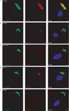
Figure 5. Mutations CCDC40 affect localization of CCDC39 in respiratory cells
Subcellular localization of CCDC39 in respiratory epithelial cells from PCD patients carrying CCDC40 loss-of-function mutations. As control, axoneme-specific antibodies against acetylated α-tubulin (green) were used. Nuclei were stained with Hoechst 33342 (blue). In respiratory epithelial cells from healthy probands (a), CCDC39 (red) localizes along the entire length of the axonemes and to a weaker degree in the apical cytoplasm. In respiratory epithelial cells from patients carrying CCDC40 loss-of function mutations OP-799 (b), OP-712 II1 (c), OP-741 (d) and OP-659 (e) CCDC39 is either markedly reduced or absent in ciliary axonemes and instead accumulates at the ciliary base. White scale bars (a–e) are 5µm.

Comment in

  • Coiled-coils and motile cilia.
    Satir P. Satir P. Nat Genet. 2011 Jan;43(1):10-1. doi: 10.1038/ng0111-10. Nat Genet. 2011. PMID: 21217638 No abstract available.

References

    1. Fliegauf M, Benzing T, Omran H. When cilia go bad: cilia defects and ciliopathies. Nat Rev Mol Cell Biol. 2007;8:880–893. - PubMed
    1. Barbato A, et al. Primary ciliary dyskinesia: a consensus statement on diagnostic and treatment approaches in children. Eur Respir J. 2009;34:1264–1276. - PubMed
    1. Duquesnoy P, et al. Loss-of-function mutations in the human ortholog of Chlamydomonas reinhardtii ODA7 disrupt dynein arm assembly and cause primary ciliary dyskinesia. Am J Hum Genet. 2009;85:890–896. - PMC - PubMed
    1. Loges NT, et al. Deletions and point mutations of LRRC50 cause primary ciliary dyskinesia due to dynein arm defects. Am J Hum Genet. 2009;85:883–889. - PMC - PubMed
    1. Omran H, et al. Ktu/PF13 is required for cytoplasmic pre-assembly of axonemal dyneins. Nature. 2008;456:611–616. - PMC - PubMed

Publication types