. 2011 Jan;18(1):49-55.
doi: 10.1038/nsmb.1948. Epub 2010 Dec 5.

β₂-microglobulin forms three-dimensional domain-swapped amyloid fibrils with disulfide linkages

Affiliations

β₂-microglobulin forms three-dimensional domain-swapped amyloid fibrils with disulfide linkages

Cong Liu et al. Nat Struct Mol Biol. 2011 Jan.

Abstract

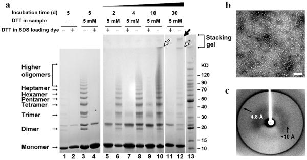
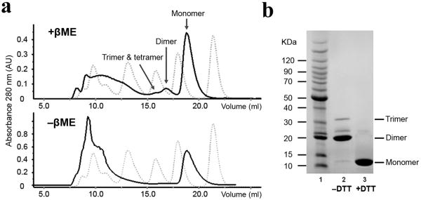
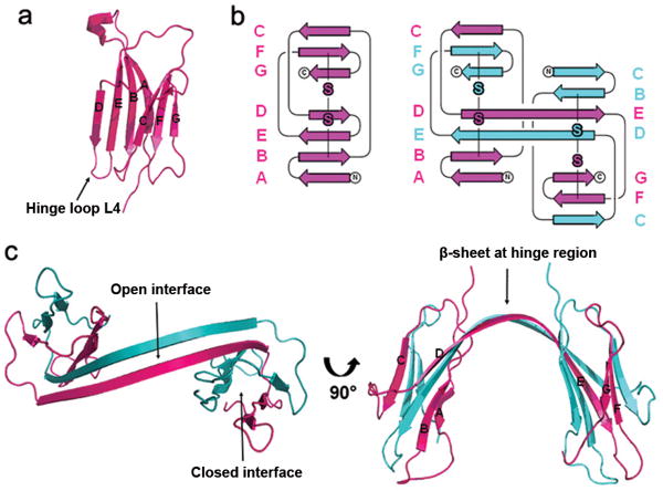
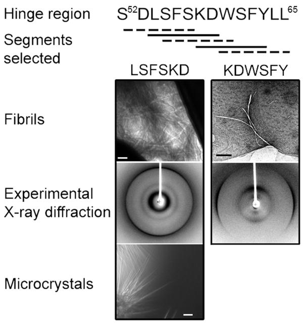
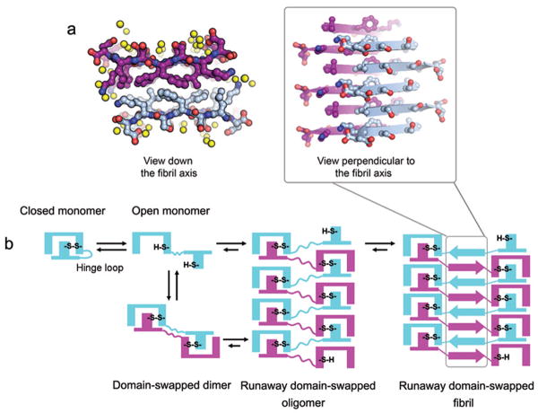
β₂-microglobulin (β₂m) is the light chain of the type I major histocompatibility complex. It deposits as amyloid fibrils within joints during long-term hemodialysis treatment. Despite the devastating effects of dialysis-related amyloidosis, full understanding of how fibrils form from soluble β₂m remains elusive. Here we show that β₂m can oligomerize and fibrillize via three-dimensional domain swapping. Isolating a covalently bound, domain-swapped dimer from β₂m oligomers on the pathway to fibrils, we were able to determine its crystal structure. The hinge loop that connects the swapped domain to the core domain includes the fibrillizing segment LSFSKD, whose atomic structure we also determined. The LSFSKD structure reveals a class 5 steric zipper, akin to other amyloid spines. The structures of the dimer and the zipper spine fit well into an atomic model for this fibrillar form of β₂m, which assembles slowly under physiological conditions.


Figures

Figure 1
Figure 1
Characterization of β2m oligomers. (a) SDS-PAGE of β2m oligomerization under non-reducing and reducing conditions. Oligomerization started from monomeric β2m with/without DTT agitated at 37°C for various times. After 5 days, without DTT, oligomer ladders still formed (lane 1), but much slower than with DTT (lane 3). Over time, the disulfide-bridged oligomer ladders were observed (lane 6, 8, 10, and 12). After 10 days, even higher oligomers formed and remained at the boundary between the stacking gel and running gel (lane 10 and 12, open arrows). After 30 days, protofilaments formed and were stuck in the loading well (lane 12, filled arrow). The oligomers were dissociated into monomers upon reduction in SDS loading buffer (+DTT) (lane 2, 4, 5, 7, 9, and 11). Lane 13 is the BenchMark Protein Ladder (Invitrogen). The additional band above the dimer which exists in all β2m samples is DTT-resistant unlike other oligomeric species. It is composed of β2m according to amino acid sequencing while mechanism of its formation remains to be determined. (b) Electron micrograph showing the protofilaments formed in (a). The scale bar is 200 nm. (c) Experimental fibril X-ray diffraction pattern of protofilaments visualized in (b).
Figure 2
Figure 2
Refolding and purification of β2m dimer. (a) Analytical size-exclusion chromatography elution profiles of β2m after refolding. The dotted profiles show five molecular weight markers (Bio-Rad gel filtration standard). From right to left: vitamin B12 (Mr 1,350), equine myoglobin (Mr 17,000), chicken ovalbumin (Mr 44,000), bovine gamma globulin (Mr 158,000) and thyroglobulin (Mr 670,000). The solid profiles show β2m refolding from inclusion bodies with/without βME. (b) Purified dimer on SDS-PAGE. Lane 1, BenchMark Protein Ladder; lane 2, 3, β2m dimer in the absence/presence of DTT in SDS loading dye.
Figure 3
Figure 3
Structure of the domain-swapped β2m dimer. (a) Ribbon diagram of the crystal structure of monomeric β2m (PDB entry 1LDS34). (b) Topology diagrams of β2m monomer and dimer. Intra- and intermolecular disulfide bonds are highlighted in the same color as the backbones. (c) Ribbon diagram of the crystal structure of β2m domain-swapped dimer.
Figure 4
Figure 4
Systematic screening for amyloidogenic segments in the hinge loop (residues 52-65). Five segments were selected and synthesized. LSFSKD and KDWSFY in solid lines formed fibrils, and LSFSKD also formed microcrystals. The scale bars are 100 nm for electron microscopy (fibrils) and 50 μm for light microscopy (microcrystals). The experimental X-ray diffraction images show a typical cross-β fibril diffraction pattern.
Figure 5
Figure 5
Crystal structure of segment LSFSKD and schematics for β2m fibrillation. (a) Atomic structure of hinge loop segment LSFSKD. Yellow spheres represent water molecules. The backbone of one sheet is magenta, and the backbone of the other sheet is blue. The interdigitated side chains between adjacent β-sheets form a dry interface, as shown by the projection down the fibril axis on the left. The LSFSKD structure is a typical steric zipper structure, belonging to Class 5. (b) Schematic model for β2m fibrillation via domain swapping. Upon reduction of the intramolecular disulfide bond, the β2m monomer can assemble to ‘closed-ended’ oligomers, such as the dimer characterized in this work, or ‘open-ended’ runaway domain-swapped oligomers. Each subunit is colored in either blue or magenta. The formation of intermolecular disulfide bonds stabilizes the domain-swapped oligomers. The self-association of hinge loops into a zipper spine accomplishes the transformation from oligomers into fibrils.

Similar articles

Cited by

References

    1. Westermark P, et al. Amyloid: toward terminology clarification. Report from the Nomenclature Committee of the International Society of Amyloidosis. Amyloid. 2005;12:1–4. - PubMed
    1. Chiti F, Dobson CM. Protein misfolding, functional amyloid, and human disease. Annu Rev Biochem. 2006;75:333–66. - PubMed
    1. Makin OS, Serpell LC. Structures for amyloid fibrils. Febs J. 2005;272:5950–61. - PubMed
    1. Nelson R, et al. Structure of the cross-β spine of amyloid-like fibrils. Nature. 2005;435:773–8. - PMC - PubMed
    1. Sawaya MR, et al. Atomic structures of amyloid cross-β spines reveal varied steric zippers. Nature. 2007;447:453–7. - PubMed

Publication types

LinkOut - more resources