

Architecture Is the Message: The role of extracellular matrix and 3-D structure in tissue-specific gene expression and breast cancer
- PMID: 21132084
- PMCID: PMC2995891
Item in Clipboard
Architecture Is the Message: The role of extracellular matrix and 3-D structure in tissue-specific gene expression and breast cancer
Pezcoller Found J.
2007 Oct.
Abstract
I was honored to deliver the 2(nd) Stanley Korsmeyer memorial Lecture on May 9(th), 2007 in Padova, Italy. Stan will always occupy a very special place in my heart: I admired him greatly not only for his magnificent and original science but also for his integrity and his grace. This review, which summarizes my laboratory's contribution to cell and cancer biology in the last 30 years, is dedicated to Stan's memory, and to Elaine Fuchs, one of my most cherished friends without whose support this work would not have gained the degree of recognition it enjoys today. My thanks also to the Pezcoller Foundation for making that week in May, 2007 one of the most memorable in my scientific life.
Figures
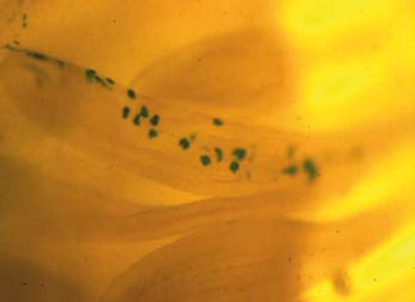
Contribution of v-Src-infected cells to normal tissue structures during chick embryo development. Chick limb buds were infected at day 4 in ovo (embryonic stage 24) with a virus encoding v-Src and a genetic marker, beta-galactosidase. The contribution of v-Src-infected cells to normal tissues (in this case a day 14 feather filament) is revealed by X-gal staining of embryo whole mounts (unpublished picture: A. Stoker and M.J.Bissell, unpublished photomicrograph: see (12). Reproduced also in (13).
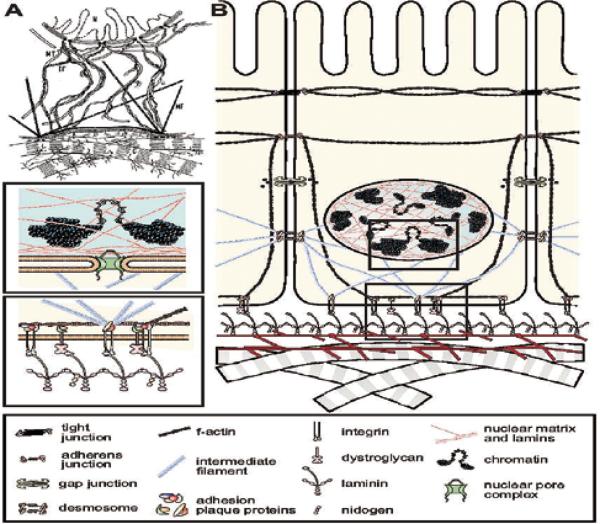
(A) Two versions of the model of dynamic reciprocity. The original model of dynamic reciprocity, or “the minimum required unit for tissue-specific functions”. N=nucleus; MT=microtubules; IF=intermediate filaments; MF=microfilaments; C=collagen. Reproduced with permission from (8) (B) An updated view of dynamic reciprocity. Reproduced with permission from (3).
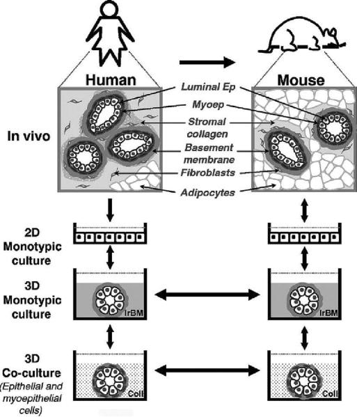
Hierarchical modeling of human breast function. Similarities between the organization of human and mouse mammary glands have enabled our observations in one tissue to be transferred to the other(keeping in mind that they are not identical in all aspects). This dynamic exchange of information has led to the gradual development of mammary gland models that now represent a continuum of organotypic systems ranging in complexity from monotypic 3D cultures to multicellular co-cultures to in vivo xenograft models. Each of the 3D models depicted here represents a physiologically relevant assay in its own right. However, when engineered with common cellular components and used in series, these models become invaluable tools for the identification and verification of disease-related molecules as well as for the design and translation of effective drug therapies. Future models that are more faithful to the human mammary microenvironment may be achieved by adding other cell types that interact within the mammary gland: fibroblasts, endothelial and fat cells as well as immune cells such as mast cells. Ep, epithelial cell; Myoep, myoepithelial cell. Adapted from previous publications and produced with permission from (–55).
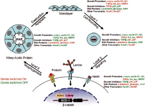
An illustration of the different levels through which ECM controls gene expression and tissue function. As cells transition from a 2D monolayer to a 3D environment, they undergo changes in cell shape that influence the expression of certain genes. Exposure to ECM engages specific cell surface receptors and initiates the transduction of biochemical and mechanical signals through the cell to the nucleus, where they further influence gene expression. As the duration of exposure time to ECM increases, cells undergo morphogenic events involving the formation of acinar structures, and once again exhibit changes in their gene expression profile. Thus, tissue structure influences gene expression and, therefore, dictates tissue function. (Modified, with permission, from 56,10,2).
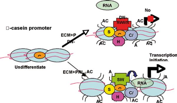
Model displaying how exposure of mammary epithelial cells to ECM and prolactin may induce the recruitment of transcription factors and chromatin remodeling enzymes to the -casein promoter, and how aberrations in SWI/SNF function interfere with RNA polymerase II recruitment.(Reproduced with permission from 32).
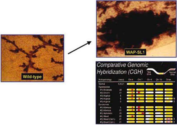
MMP 3 overexpression leads to formation of mammary tumors as mice age: A mutated form of MMP3 (SL-1) was attached to the promoter of the whey acidic protein gene to engineer the mouse. This milk protein begins to be expressed essentially on midpregnancy and this constructs effectively delivers a large amount of MMP3 essentially to the mammary gland of pregnant mice. Animals develop mammary tumors as they age and the tumors are shown to have substantial genomic defects measured by Comparative Genomic Hybridization (CGH). Adopted from (36).
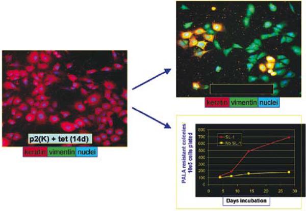
MMP3 causes EMT and genomic instability in ScP2 cells: Mouse mammary epithelial cells (mutant for p53) were transfected with a tet repressible MMP3 (the same mutated MMP3 used in Fig.6; 35). Removal of tet led to expression of MMP3, changes in the cytoskeleton, EMT and genomic instability measured by amplification of the CAD locus. Adapted from (39, 41).
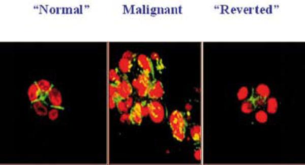
1-inhibitory antibody treatment of tumor cells leads to the formation of reverted acini. Confocal fluorescence microscopy images of F actin: Both the S-1 (left) and T4-2 - reverted acini (right) showed basally localized nuclei (propidium iodide) and organized filamentous F-actin (FITC), while T4-2 mock-treated colonies (T4-2 IgG, middle) had disorganized, hatched bundles of actin and pleiomorphic nuclei. Adapted from previous publication (Reproduced with permission from 43).
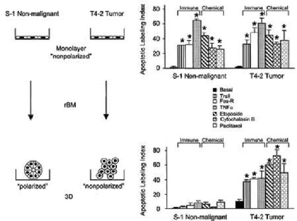
Schematic presentation of data obtained on the importance of 3D structure and tissue polarity in response of non malignant and malignant breast cancer cell lines to chemotherapy Only nonmalignant cells within an organized and polarmammary acini are resistant to apoptosis. But when T4-2 cells are `reverted' to an organized structure in 3D (see section VII), they too become resistant, These findings have profound implication for dormancy in breast cancer. Apoptotic labeling indices calculated for S-1 and T4-2 cells treated with Trai. Modified from (46).
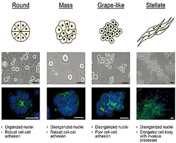
Breast cell line colony morphologies in 3D culture fall into four distinct groups. A panel of twenty-five breast cell lines were cultured in three-dimensions and grouped into four distinct morphologies. A schematic and key descriptors of each morphology is shown on top in addition to phase contrast (middle) and F-actin and nuclear fluorescence images(bottom) of representative cell lines of each morphology. Scale bars: phase contrast, 50 μm; fluorescence, 20 μm. Reproduced with permission from (49)
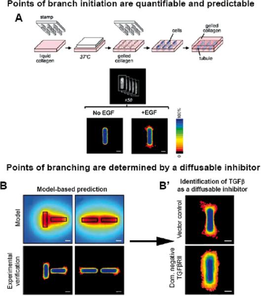
(A) Schematic of 3D microfabrication method to engineer tubules. The position of cells was quantified by stacking images of nuclei from 50 tubules to generate a frequency map before induction of branching. F frequency map of tubules 24 hours after adding EGF to induce branching. B. Position of branching can be predicted by calculated concentration profile. Calculated profiles of diffusible inhibitors in tubules oriented perpendicular and parallel to each other. Frequency maps 24 hours after induction of branching confirm that branching is inhibited in regions predicted to be surrounded by a high concentration of inhibitors in perpendicular and parallel tubules. Scale bars, 50 mm. B' Positional control of branching is disrupted by blocking signaling of endogenous TGF 1. Shown are frequency maps 24 hours after induction of branching in tubules of vector control cells and (E) cells overexpressing dominant negative TGFb receptor type II (HA-DNTbRII). Scale bars, 50 mm. Reproduced with permission from (51).
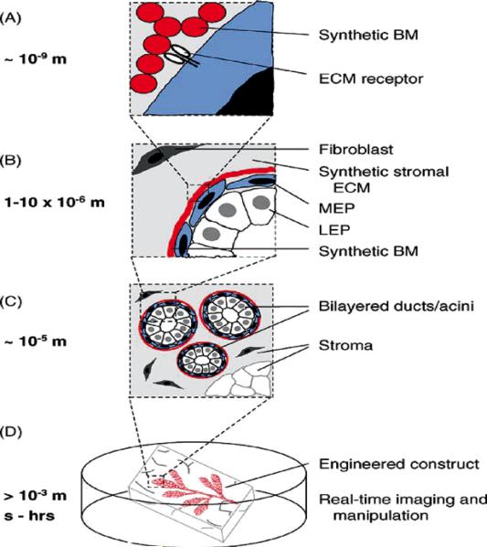
The tissue-engineered breast. New strategies should enable the control of the microenvironment at the nano-, micro-, and macro-scales with temporal precision. (A) Synthetic and recombinant ECM polymers impart cues sensed directly by cell-surface receptors. (B–C) Microfabricated constructs control the positions of multiple cell types with respect to each other and the ECM with micrometer precision across large areas of tissue. (D) Engineered breast tissues that can be visualized and manipulated in real time. Adapted from previous publication (Reproduced with permission from 52).
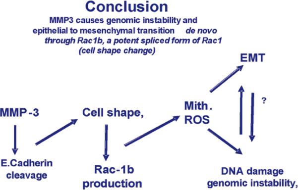
Flow chart of the steps postulated to give rise to genomic instability and EMT as a result of MMP3 activation, Adapted from previous publication (41).
References
Grants and funding
LinkOut - more resources
Full Text Sources