. 2010 Nov 15;10(12):5007-5019.
doi: 10.1021/cg1011633.

Nucleation

Affiliations

Nucleation

Peter G Vekilov. Cryst Growth Des. .

Abstract

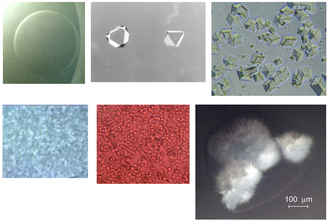
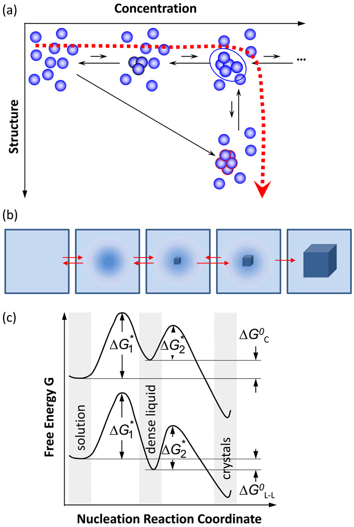
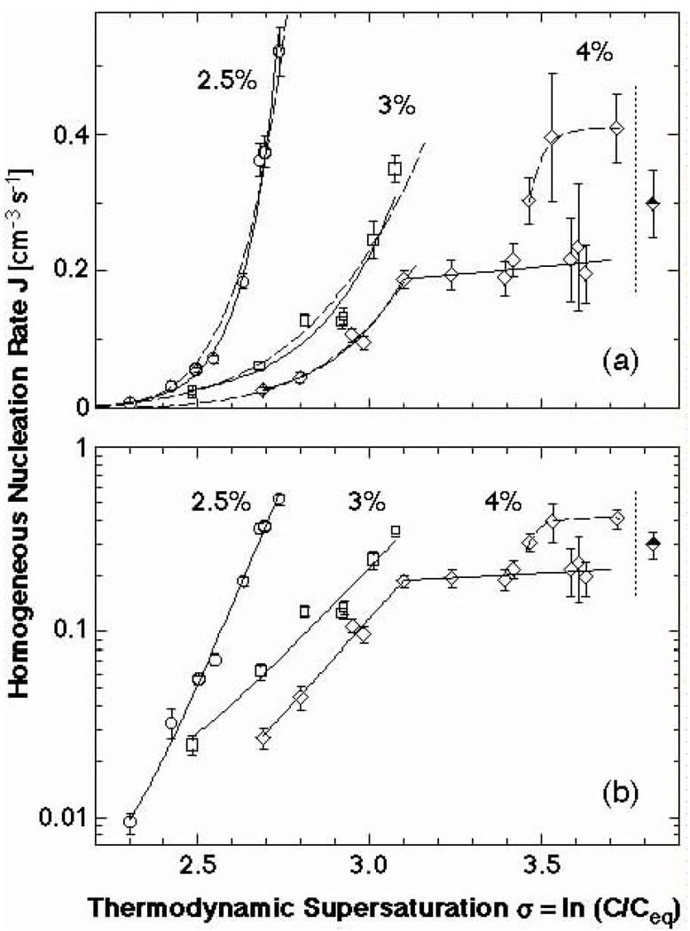
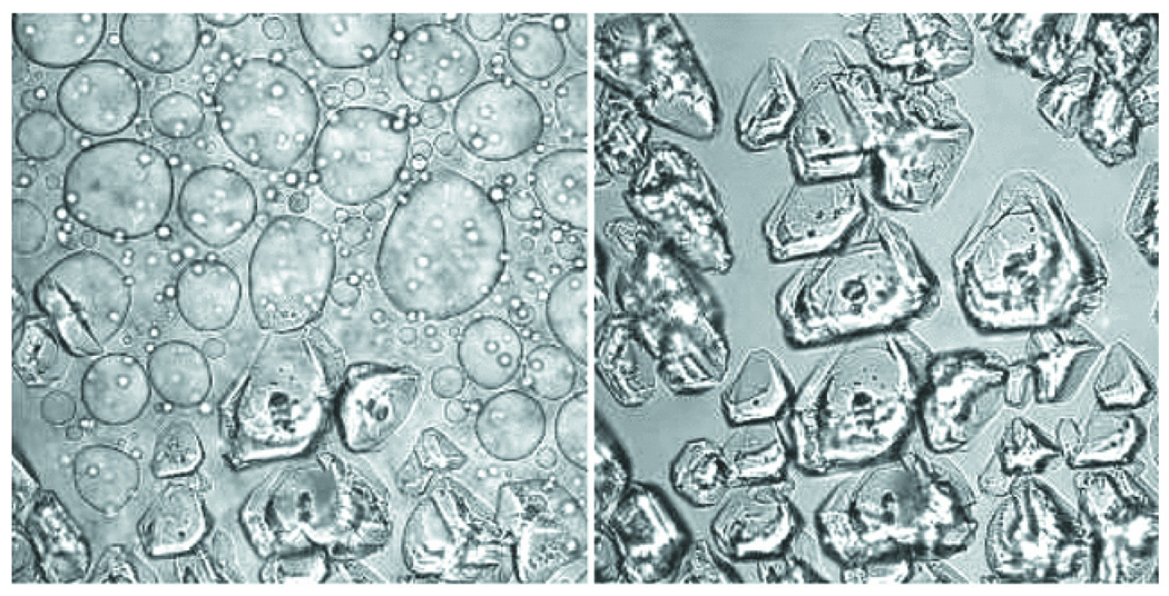
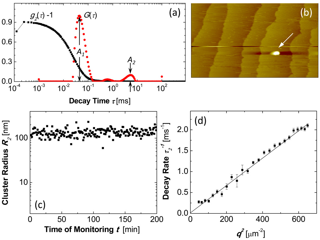
Crystallization starts with nucleation and control of nucleation is crucial for the control of the number, size, perfection, polymorphism and other characteristics of crystalline materials. This is particularly true for crystallization in solution, which is an essential part of processes in the chemical and pharmaceutical industries and a major step in physiological and pathological phenomena. There have been significant recent advances in the understanding of the mechanism of nucleation of crystals in solution. The foremost of these are the two-step mechanism of nucleation and the notion of the solution-crystal spinodal. According to the two-step mechanism, the crystalline nucleus appears inside pre-existing metastable clusters of size several hundred nanometers, which consist of dense liquid and are suspended in the solution. While initially proposed for protein crystals, the applicability of this mechanism has been demonstrated for small molecule organic materials, colloids, polymers, and biominerals. This mechanism helps to explain several long-standing puzzles of crystal nucleation in solution: nucleation rates which are many orders of magnitude lower than theoretical predictions, the significance of the dense protein liquid, and others. At high supersaturations typical of most crystallizing systems, the generation of crystal embryos occurs in the spinodal regime, where the nucleation barrier is negligible. The solution-crystal spinodal helps to understand the role of heterogeneous substrates in nucleation and the selection of crystalline polymorphs. Importantly, these ideas provide powerful tools for control of the nucleation process by varying the solution thermodynamic parameters.


Figures

Figure 1
Figure 1
Nucleation largely determines the outcome of crystallization. Examples of protein crystals and other condensed phases illustrate, at top left, the failure of nucleation, where no crystals or other condensed phase is generated in a supersaturated lysozyme solution; and clockwise from there, the nucleation of two crystals of apoferritin, which grow to a relatively large size; the nucleation of numerous crystals of insulin, which have a broad size distribution; needle-like crystals of lysozyme; dense liquid droplets in a solution of hemoglobin A, and, at bottom left, amorphous precipitate in a supersaturated lysozyme solution. Scale bar is shown in bottom right panel.
Figure 2
Figure 2
Illustration of the thermodynamic effects of formation of a crystal. n – number of molecules in crystalline embryo; Δμ – solution supersaturation; α – surface free energy; ΔG – free energy; * denotes critical cluster.
Figure 3
Figure 3
Schematic illustration of the two-step mechanism of nucleation of crystals. A dense liquid cluster forms. A crystal nucleus may form inside the cluster. (a) Microscopic viewpoint in the (Concentration, Structure) plane; (b) Macroscopic viewpoint of events along thick dashed line in (a). (c) The free-energy ΔG along two possible versions of the two step nucleation mechanism. If dense liquid is unstable and ΔGLL0>0 (ΔGLL0 —standard free energy of formation of dense liquid phase), dense liquid exists as mesoscopic clusters, ΔGLL0 transforms to ΔGC0, and upper curve applies; if dense liquid is stable, ΔGLL0<0, reflected by lower curve. ΔG1* is the barrier for formation of a cluster of dense liquid, ΔG2* – for a formation of a crystalline nucleus inside the dense liquid.
Figure 4
Figure 4
The dependence of the rate of homogeneous nucleation J of lysozyme crystals of supersaturation σ ≡ Δμ/kBT at T = 12.6 °C and at the three concentrations of the precipitant NaCl indicated on the plots. Solid lines – fits with exponential functions; dashed lines fits with the classical nucleation theory expression, Eq. (3). Vertical dotted lines at σ = 3.9 indicate the liquid-liquid coexistence boundary at this T and CNaCl = 4 %; this supersaturation corresponds to lysozyme concentration 67 mg ml−1. (a) Linear coordinates; (b) semi-logarithmic coordinates. With permission from Ref. .
Figure 5
Figure 5
The dependence of the rate of homogeneous nucleation J of lysozyme crystals on temperature T at two fixed lysozyme concentrations indicated in the plot. The temperatures of equilibrium between crystals and solution are 315 K at Clys = 50 mg ml−1 and 319 K at Clys = 80 mg ml−1. The temperatures of L-L separation are 285 K at Clys = 50 mg ml−1 and 287 K at Clys = 80 mg ml−1 and are marked with vertical dashed lines. Symbols represent experimental results from . Lines are results of two-step model in Eqs. (5)–(7). With permission from Ref. .
Figure 6
Figure 6
The phase diagram of a lysozyme solution determined experimentally in 0.05 M Na acetate buffer at pH = 4.5 and 4.0 % NaCl. Liquidus, or solubility lines—from Refs. –, liquid-liquid (L-L) coexistence and respective spinodal—from Ref. , gelation line—from Refs. –. Solution-crystal spinodal is highlighted in red and is from Ref. .
Figure 7
Figure 7
Confocal scanning laser fluorescence microscopy imaging of nucleation of crystals of glucose isomerase within dense liquid droplets. Bright field imaging, polyethylene glycol with molecules mass 10,000 g mol−1 (PEG 10000) used to induce crystallization. The time interval between the left and right images is 380 s. Cprotein = 55 mg ml−1, CPEG = 9.5%, 0.5 M NaCl, 10 mM Tris maintaining pH = 7. The width of each image is 326 µm. With permission from Ref. .
Figure 8
Figure 8
Characterization of dense liquid clusters. (a) Examples of correlation function of the scattered intensity g2(τ) and the respective intensity distribution function G(τ) of a lysozyme solution with C = 148 mg ml−1 in 20 mM HEPES buffer; data collected at angle 145°. (b) Atomic force microscopy imaging of liquid cluster landing on the surface of a crystal in a lumazine synthase solution. Tapping mode AFM imaging, scan width 20 µm. Apparent lateral cluster dimensions are misleading, cluster height is 120 nm, with permission from Ref. . (c) Time dependence of the radius of dense liquid clusters in the same lysozyme solution as in a. (d) The dependence of the decay rate Γ2 = τ2−1 of the cluster peak in the correlation function on the squared wave vector q2 for a lysozyme solution as in (a).

References

    1. De Yoreo JJ, Burnham AK, Whitman PK. Developing KH2PO4 and KD2PO4 crystals for the world's most power laser. International Materials Reviews. 2002;47(3):113–152.
    1. Reichert P, McNemar C, Nagabhushan N, Nagabhushan TL, Tindal S, Hruza A. Metal-interferon-alpha crystals. 1995;5:441. 734.
    1. Brange J. Galenics of Insulin. Berlin: Springer; 1987. ed.
    1. Long ML, Bishop JB, Nagabhushan TL, Reichert P, Smith GD, DeLucas LJ. Protein crystal growth in microgravity review of large scale temperature induction method. J. Crystal Growth. 1996;168:233–243.
    1. Matsuda S, Senda T, Itoh S, Kawano G, Mizuno H, Mitsui Y. New crystal form of recombinant murine interferon-β. J. Biol. Chem. 1989;264:13381–13382. - PubMed

LinkOut - more resources