. 2010 Mar 1;3(1):76-83.
doi: 10.1007/s12195-010-0106-2.

Rho Kinase Regulation of Fibroblast Migratory Mechanics in Fibrillar Collagen Matrices

Affiliations

Rho Kinase Regulation of Fibroblast Migratory Mechanics in Fibrillar Collagen Matrices

Chengxin Zhou et al. Cell Mol Bioeng. .

Abstract

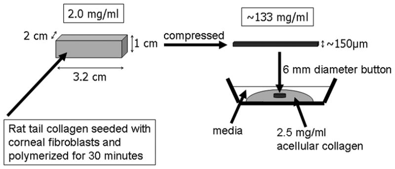
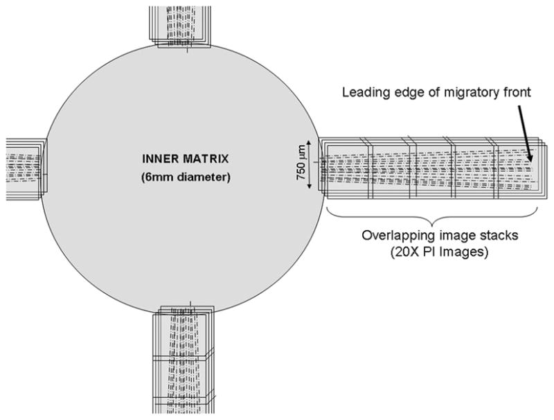
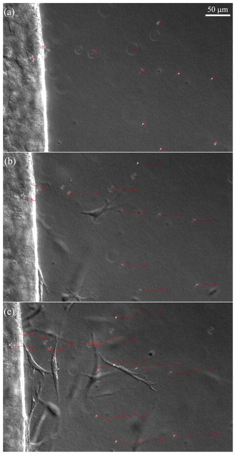
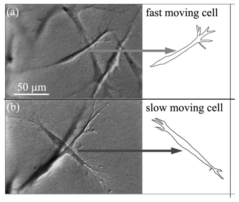
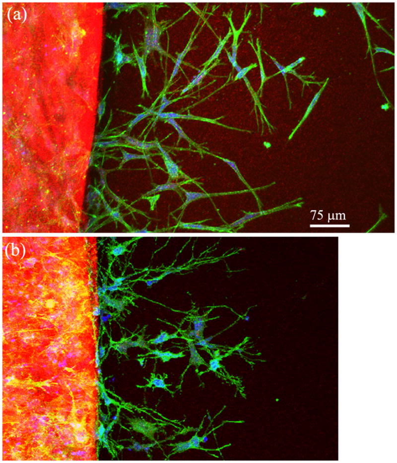
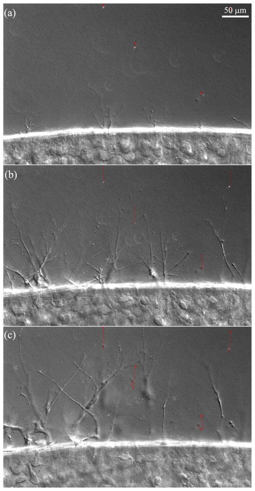
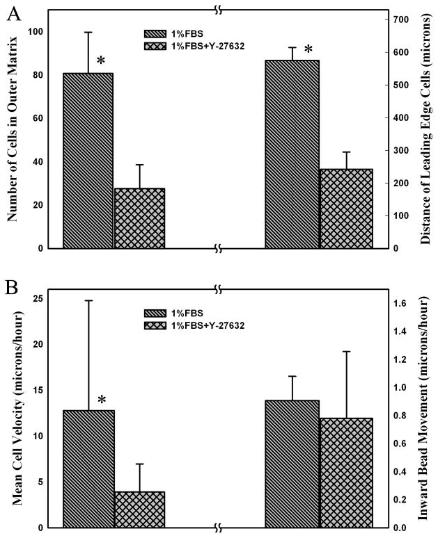
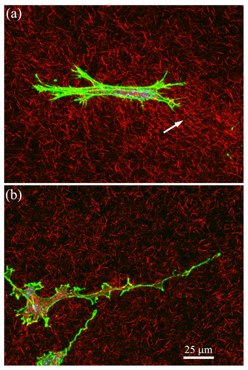
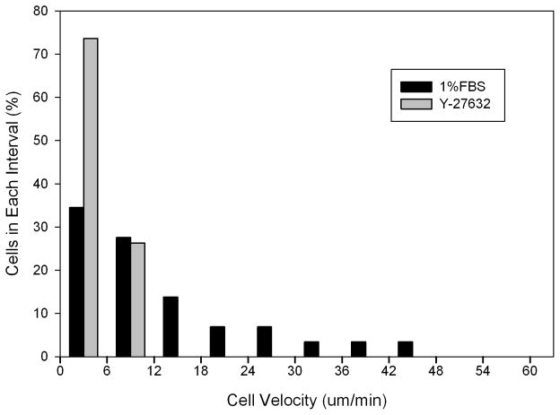
Migration of activated corneal fibroblasts plays an important role in matrix patterning during embryonic development and wound repopulation following injury or refractive surgery. In this study, we investigate the role of Rho kinase in regulating fibroblast migration mechanics, by modifying a previously described nested collagen matrix model to facilitate dynamic imaging of cell-matrix interactions.Human corneal fibroblasts were cultured in nested matrices with media containing either 1% fetal bovine serum (FBS), or 1% FBS plus the Rho kinase inhibitor Y-27632. Time-lapse DIC imaging of cell and extracellular matrix (ECM) movements was performed for up to 72 hours. In addition, static confocal imaging was used to assess 3-D cell morphology and local matrix reorganization.In 1% FBS, significant tractional forces were generated during migration, as indicated by inward displacement and reorganization of collagen in front of cells. When Rho kinase was inhibited, cells became more elongated, and extended dendritic processes into the outer matrix. Interestingly, these dendritic cells were still able to generate tractional forces at their leading edge, whereas cell translocation was substantially reduced. Overall, the data suggests that Rho kinase impacts 3-D fibroblast migration by affecting morphology, polarization, and mechanical coordination between the leading and trailing edges of cells.


Figures

Figure 1
Figure 1
Method for constructing nested matrix model. (Modified from Kim A, Lakshman N, Karamichos D, Petroll WM. Growth factor regulation of corneal keratocyte differentiation and migration in compressed collagen matrices. Invest Ophthalmol Vis Sci)
Figure 2
Figure 2
Schematic showing the montage of 3-D image stacks collected in each sample using laser scanning confocal microscopy. (reproduced from: Karamichos D, Lakshman N, Petroll WM. An Experimental Model for Assessing Fibroblast Migration in 3-D Collagen Matrices. Cell Motil Cytoskeleton 66(1):1–9, Copyright © 2009 Wiley-Blackwell)
Figure 3
Figure 3
Time-lapse DIC images collected at 6 hours, 24hours and 48 hours from an experiment in which cells were cultured in 1% FBS. Red tracks show movement of embedded microspheres (crosses mark starting positions). Note that as fibroblasts move out of the inner matrix (left), the beads are pulled inward.
Figure 4
Figure 4
DIC images of typical fast and slow moving cells in 1%FBS. (A) Fast migrating cell has a clear, persistent leading edge with pseudopodia, and a streamline tail. (B) Slow migrating cell has less distinctive leading and rear edges. Both the front and rear have appreciable pseudopodia.
Figure 5
Figure 5
20X confocal maximum intensity projection images of f-actin (green), collagen fibrils (red) and PI (blue) following 72 hours of culture in (A) 1% FBS, or (B) Y-27632 (10 μM).
Figure 6
Figure 6
Time-lapse DIC images collected at 6 hours, 24hours and 48 hours from an experiment in which cells were cultured in 1% FBS + Y-27632. Red tracks show movement of embedded microspheres (crosses mark starting positions). Note that as fibroblasts move out of the inner matrix (bottom), the beads are pulled inward a substantial distance. However, cells assume a more dendritic morphology following Rho kinase inhibition (compare to Figure 3), and take longer to completely escape the inner matrix.
Figure 7
Figure 7
Quantitative analysis of 72 hour confocal (A) and time-lapse DIC (B) image data. (A) Comparison of number of cell in outer matrix and the distance cells traveled in 1% FBS and 1%FBS plus 10 μM Y-27632. Graphs show mean and standard deviations of 3 constructs per condition. (B) Speed of cell and bead movement under different culture conditions from time-lapse DIC imaging experiments. Inhibiting Rho kinase with Y-27632 significantly reduced the rate of cell movement, but not the net inward bead movement. (Number of cells analyzed was 29 for1% FBS and 19 for 1%FBS plus 10 μM Y-27632. Number of beads analyzed was 15 for1% FBS and 13 for 1%FBS plus 10 μM Y-27632.) * P < 0.05.
Figure 8
Figure 8
63X confocal maximum intensity projection images (10 microns thick) of f-actin (green), collagen fibrils (red) and PI (blue) after 3 days of culture. A) Migrating cell near leading edge under 1%FBS condition. Cell is bipolar, and compaction of collagen is observed at one end of the cell (arrow). B) Migrating cell near leading edge under 1%FBS + Y-27632 condition. Cell has an elongated dendritic morphology, and the surrounding ECM is less compacted.
Figure 9
Figure 9
Histograms of cell velocities under different culture conditions, from time-lapse DIC imaging experiments. Inhibiting Rho kinase with 10 μm Y-27632 dramatically reduced the range of cell velocities.

Similar articles

Cited by

References

    1. Amano M, Ito M, Kimura K, Fukata Y, Chihara K, Nakano T, Matsuura Y, Kaibuchi K. Phosphorylation and activation of myosin by Rho-associated kinase (Rho-kinase) J Biol Chem. 1996;271:20246–20249. - PubMed
    1. Brown RA, Wiseman M, Chuo CB, Cheema U, Nazhat SN. Ultrarapid engineering of biomimetic materials and tissues: Fabrication of nano- and microstructures by plastic compression. Advanced Functional Materials. 2005;15:1762–1770.
    1. Fischer DJ, Liliom K, Guo Z, Nusser N, Virag T, Murakami-Murofushi K, Kobayashi S, Erickson JR, Sun G, Miller DD, Tigyi G. Naturally occurring analogs of lysophosphatidic acid elicit different cellular responses through selective activation of multiple receptor subtypes. Mol Pharmacol. 1998;54:979–988. - PubMed
    1. Fischer RS, Gardel M, Ma X, Adelstein RS, Waterman CM. Local cortical tension by myosin II guides 3D endothelial cell branching. Curr Biol. 2009;19:260–265. - PMC - PubMed
    1. Friedl P, Wolf K. Plasticity of cell migration: a multiscale tuning model. J Cell Biol. 2009;1:1. - PMC - PubMed

LinkOut - more resources