. 2011 Mar;39(3):946-52.
doi: 10.1007/s10439-010-0219-8. Epub 2010 Dec 4.

Multifunctional FePt nanoparticles for radiation-guided targeting and imaging of cancer

Affiliations

Multifunctional FePt nanoparticles for radiation-guided targeting and imaging of cancer

Ghazal Hariri et al. Ann Biomed Eng. 2011 Mar.

Abstract

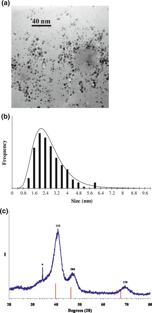
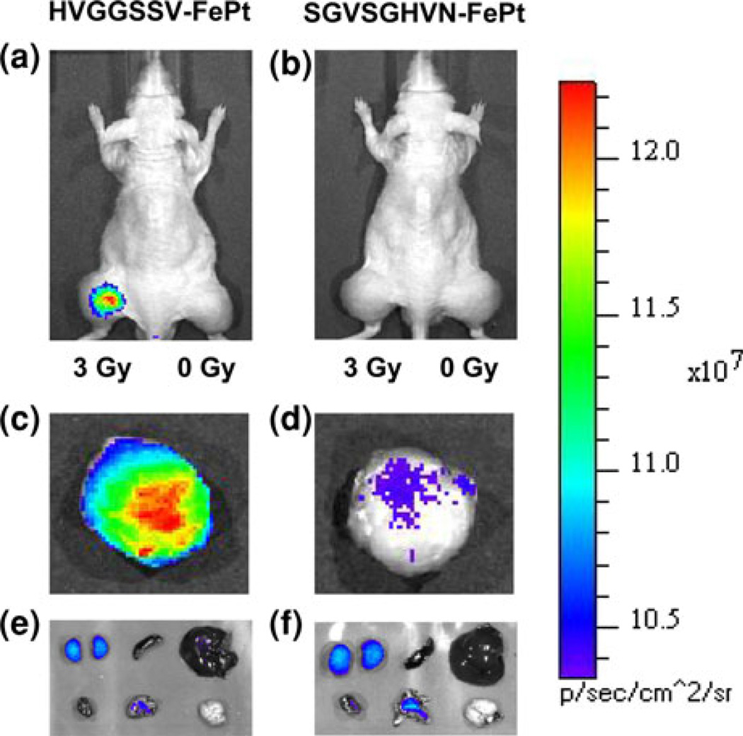
A multifunctional FePt nanoparticle was developed that targets tumor microvasculature via "radiation-guided" peptides, and is detected by both near-infrared (NIR) fluorescence imaging and analytical mass spectrometry methods. Tumor specific binding was first measured by biotinylated peptide linked to fluorophore-conjugated streptavidin. This showed tumor selective binding to tumors using the HVGGSSV peptide. FePt nanoparticles were synthesized sequentially by surface modification with poly(L)lysine, poly(ethylene) glycol conjugation, and functionalized with HVGGSSV peptide and fluorescent probe Alexa fluor 750. NIR fluorescence imaging and ICP-MS analysis showed significant HVGGSSV-FePt nanoparticle binding to irradiated tumors as compared to unirradiated tumors and controls. Results indicate that multifunctional FePt nanoparticles have potential application for radiation-guided targeting and imaging of cancer.


Figures

FIGURE 1
FIGURE 1
(a) TEM micrograph (250 kx) and particle-size histogram (b) of prepared fcc FePt nanoparticles (calculated average particle size of 2.7 ± 1.0 nm). (c) XRD scan (Cu Kα radiation) of prepared fcc FePt nanoparticles along with the XRD line pattern of Pt metal, PDF (#4-802). Peak indices are assigned relative to those known for fcc FePt. Scherrer’s analysis of XRD peak widths gives an average volume-weighted fcc FePt particle size of 2.3 nm. Asterisk denotes diffraction intensity assigned to a trace amount of Na2CO3.
FIGURE 2
FIGURE 2
In vivo NIR fluorescence imaging of subcutaneous H460 lung tumor-bearing nude mice (hind limbs) after intravenous tail vein injection of 50 µg of (a) targeted HVGGSSV-FePt nanoparticles labeled with Alexa fluor 750 and (b) scrambled SGVSGHVN-FePt nanoparticles labeled with Alexafluor 750. In both groups, the left hind limb tumor was treated with 3 Gy radiation to induce receptor expression, while the right hind limb tumor served as a control and received a sham radiation dose of 0 Gy. Images were acquired at 48 h following injection. Strong tumor binding is observed in the irradiated (left tumor) for the HVGGSSV targeted FePt nanoparticle. Ex vivo NIR fluorescence imaging of representative excised irradiated tumors at 48 h: (c) Targeted HVGGSSV-FePt nanoparticles labeled with Alexa fluor 750; (d) scrambled SGVSGHVN-FePt nanoparticles labeled with Alexa fluor 750; and internal organs (top row from left to right are kidneys, spleen, and liver; bottom row from left to right are heart, lungs, and brain). (e) Targeted HVGGSSV-FePt nanoparticles labeled with Alexa fluor 750 and (f) scrambled SGVSGHVN-FePt nanoparticles labeled with Alexa fluor 750. All NIR fluorescence images were acquired at 48 h and are normalized.
FIGURE 3
FIGURE 3
Bar graph of tumor tissue treated with 3 Gy radiation vs. normal tissues in targeted HVGGSSV-FePt nanoparticles and scrambled SGVSGHVN-FePt nanoparticles both labeled with Alexa fluor 750 (control) (n = 3–5 mice). Fluorescence intensity is expressed as units of photons/s/area/steradians. *p<0.05.
FIGURE 4
FIGURE 4
Bar graph of total platinum levels in H460 lung tumor-bearing nude mice after intravenous injection of targeted HVGGSSV-FePt nanoparticles and scrambled SGVSGHVN-FePt nanoparticles (control) via tail vein (n = 3–6 mice) *p <0.05.
SCHEME 1
SCHEME 1
Multifunctional FePt nanoparticle synthesis. Schematic diagram for synthesis of multifunctional FePt nanoparticles. (a) Surface modification of FePt-folate with poly(l)lysine, (b) addition of polyethylene glycol crosslinker, (c) conjugation of HVGGSSV peptide, and (d) conjugation of Alexafluor 750 fluorescent probe.

Similar articles

Cited by

References

    1. Babic M, Horak D, Trchova M, Jendelova P, Glogarova K, Lesny P, Herynek V, Hajek M, Sykova E. Poly(l-lysine)-modified iron oxide nanoparticles for stem cell labeling. Bioconjugate Chem. 2008;19:740–750. - PubMed
    1. Cai W, Chen X. Nanoplatforms for targeted molecular imaging in living subjects. Small. 2007;3:1840–1854. - PubMed
    1. Cai W, Shin D, Chen K, Gheysens O, Cao Q, Wang SX, Gambhir SS, Chen X. Peptide-labeled near-infrared quantum dots for imaging tumor vasculature in living subjects. Nano Lett. 2006;6:669–676. - PubMed
    1. Han Z, Fu A, Wang H, Diaz R, Geng L, Onishko H, Hallahan DE. Noninvasive assessment of cancer response to therapy. Nat. Med. 2008;14:343–349. - PubMed
    1. Hariri G, Yan H, Wang H, Han Z, Hallahan DE. Radiation-guided drug delivery to cancer. Clin. Cancer Res. 2010;16:4968–4977. - PMC - PubMed

Publication types