. 2010 Dec 6:3:328.
doi: 10.1186/1756-0500-3-328.

Use of RNAlater in fluorescence-activated cell sorting (FACS) reduces the fluorescence from GFP but not from DsRed

Affiliations

Use of RNAlater in fluorescence-activated cell sorting (FACS) reduces the fluorescence from GFP but not from DsRed

Ismail Zaitoun et al. BMC Res Notes. .

Abstract

Background: Flow cytometry utilizes signals from fluorescent markers to separate targeted cell populations for gene expression studies. However, the stress of the FACS process could change normal gene expression profiles. RNAlater could be used to stop such changes in original gene expression profiles through its ability to denature RNase and other proteins. The normal conformational structure of fluorescent proteins must be maintained in order to fluoresce. Whether or not RNAlater would affect signals from different types of intrinsic fluorescent proteins is crucial to its use in flow cytometry; this question has not been investigated in detail.

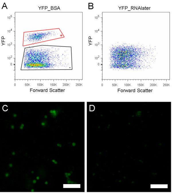
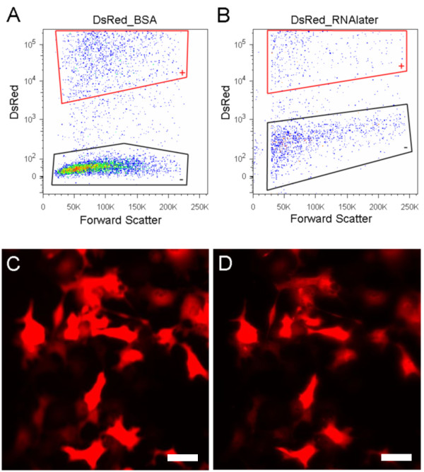
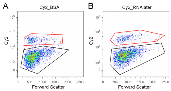
Findings: To address this question, we analyzed the effect of RNAlater on fluorescence intensity of GFP, YFP, DsRed and small fluorescent molecules attached to secondary antibodies (Cy2 and Texas-Red) when used in flow cytometry. FACS results were confirmed with fluorescence microscopy. Our results showed that exposure of YFP and GFP containing cells to RNAlater reduces the intensity of their fluorescence to such an extent that separation of such labeled cells is difficult if not impossible. In contrast, signals from DsRed2, Cy2 and Texas-Red were not affected by RNAlater treatment. In addition, the background fluorescence and clumping of dissociated cells are altered by RNAlater treatment.

Conclusions: When considering gene expression studies using cell sorting with RNAlater, DsRed is the fluorescent protein of choice while GFP/YFP have severe limitations because of their reduced fluorescence. It is necessary to examine the effects of RNAlater on signals from fluorescent markers and the physical properties (e.g., clumping) of the cells before considering its use in cell sorting.


Figures

Figure 1
Figure 1
RNAlater decreases fluorescence of YFP. (A & B) FACS results. All data in FACS figures are restricted to cells defined by forward and side light scatter and further to singlet events. The YFP positive cells are shown enclosed by red lines and negative cells are shown enclosed by black lines. (A) Cells in BSA: a population of YFP positive cells is clearly distinguished from YFP negative cells. (B) Cells in RNAlater: the two populations are not discernable. (C & D) dissociated cells were observed under the fluorescence microscope (C) YFP positive cells in BSA. (D) YFP positive cells one minute after addition of RNAlater. The intensity of the YFP decreased substantially in the presence of RNAlater. (C & D) Exposure time: 200ms; scale bar: 50 microns.
Figure 2
Figure 2
Fluorescence of DsRed2 protein is not affected in RNAlater. (A & B) FACS results. All data in FACS figures are restricted to single cells defined by forward and side light scatter; clumped and ruptured cells (debris) are not displayed in the figure. DsRed positive and negative COS-7 cells were mixed before flow cytometry sorting. (A) Cells in BSA: a population of DsRed2 positive cells is clearly distinguished from DsRed2 negative cells. The cells with intermediate fluorescence intensity between the positive and negative populations represent newly dividing cells, which are in the initial stages of DsRed expression. (B) Cells in RNAlater: fewer cells are shown here than in (A) because RNAlater has induced cell clumping, so fewer singlets are available to the sorter. RNAlater did not quench fluorescent signals from analyzed DsRed positive cells. (C & D) Dissociated cells were observed under the fluorescence microscope (C) DsRed2 positive cells in BSA. (D) DsRed2 positive cells after addition of RNAlater. The intensity of the DsRed2 was stable in the presence of RNAlater. (C & D) Exposure time: 30ms; scale bar: 50 microns.
Figure 3
Figure 3
Fluorescence of Cy2 is not affected in RNAlater. (A & B) FACS results. All data in FACS figures are restricted to cells defined by forward and side light scatter and further to singlet events. Dissociated cells were fixed and immunostained. The immunostained cells were visualized with secondary antibody conjugated with Cy2. The Cy2 positive cells are shown enclosed by red lines and negative cells are shown enclosed by black lines. (A) Cells in BSA: a population of Cy2 positive cells is clearly distinguished from Cy2 negative cells. (B) Cells in RNAlater: the two populations are distinguishable as well.

References

    1. Alenghat FJ, Ingber DE. Mechanotransduction: all signals point to cytoskeleton, matrix, and integrins. Sci STKE. 2002;2002:pe6. doi: 10.1126/stke.2002.119.pe6. - DOI - PubMed
    1. Avvisato CL, Yang X, Shah S, Hoxter B, Li W, Gaynor R, Pestell R, Tozeren A, Byers SW. Mechanical force modulates global gene expression and beta-catenin signaling in colon cancer cells. J Cell Sci. 2007;120(15):2672–2682. doi: 10.1242/jcs.03476. - DOI - PubMed
    1. Maxwell WMC, Welch GR, Johnson LA. Viability and membrane integrity of spermatozoa after dilution and flow cytometric sorting in the presence or absence of seminal plasma. Reprod Fertil Dev. 1997;8:1165–1178. doi: 10.1071/RD9961165. - DOI - PubMed
    1. Seidl J, Knuechel R, Kunz-Schughart LA. Evaluation of membrane physiology following fluorescence activated or magnetic cell separation. Cytometry. 1999;36:102–111. doi: 10.1002/(SICI)1097-0320(19990601)36:2<102::AID-CYTO3>3.0.CO;2-D. - DOI - PubMed
    1. Ma N, Koelling K, Chalmers JJ. The fabrication and use of a transient contractional flow device to quantify the sensitivity of mammalian and insect cells to hydrodynamic forces. Biotechnol Bioeng. 2002;80:428–437. doi: 10.1002/bit.10387. - DOI - PubMed

LinkOut - more resources