. 2010 Dec 6:7:89.
doi: 10.1186/1742-2094-7-89.

Microglial activation state exerts a biphasic influence on brain endothelial cell proliferation by regulating the balance of TNF and TGF-β1

Affiliations

Microglial activation state exerts a biphasic influence on brain endothelial cell proliferation by regulating the balance of TNF and TGF-β1

Jennifer V Welser et al. J Neuroinflammation. .

Abstract

Background: Studies of cerebral ischemia and other neuroinflammatory states have demonstrated a strong association between new vessel formation and microglial recruitment and activation, raising the possibility that microglia may be involved in promoting angiogenesis. As endothelial cell proliferation is a fundamental early step in angiogenesis, the aim of this study was to test this hypothesis by examining the influence of microglial secreted factors on brain endothelial cell (BEC) proliferation using BrdU incorporation.

Methods: Primary cultures of mouse BEC, microglia and astrocytes were used in this study. Proliferation of BEC was examined by BrdU incorporation. ELISA was used to quantify TNF and TGF-β1 levels within cell culture supernatants.

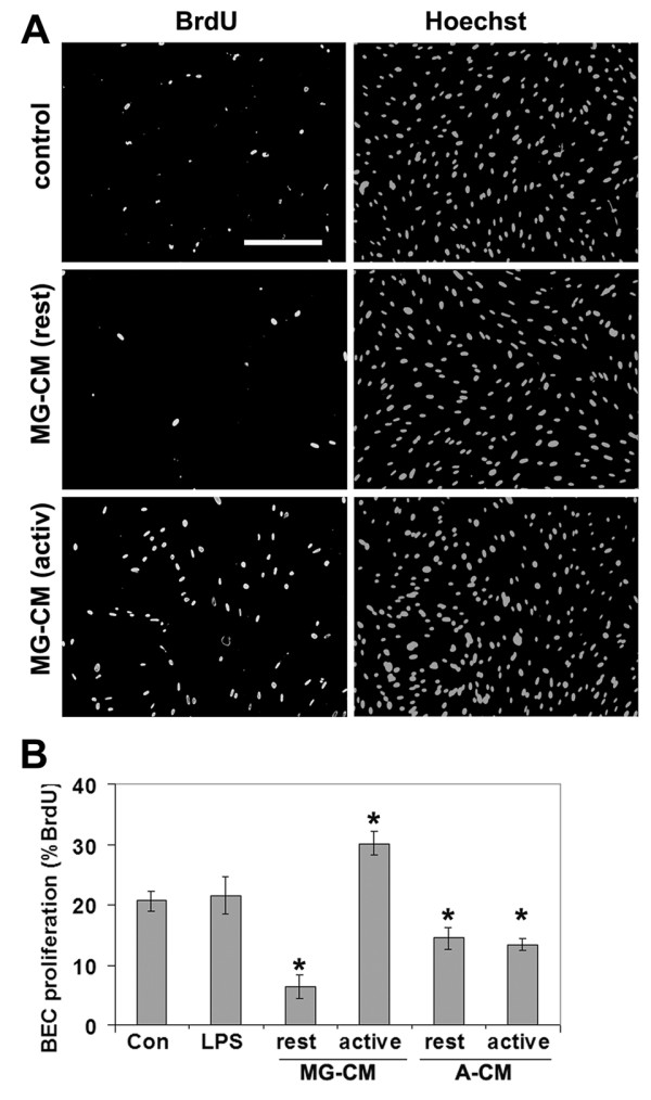
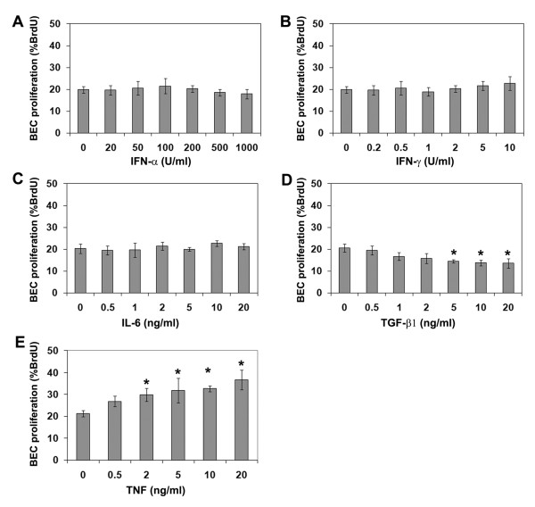
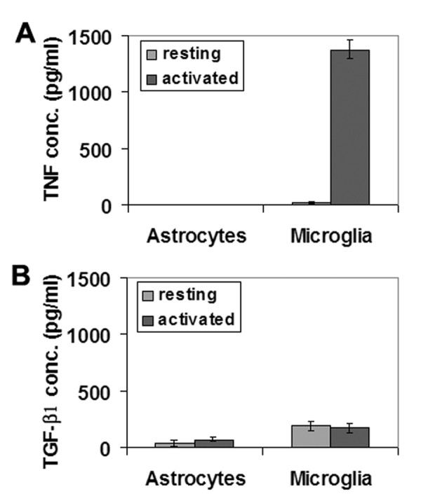
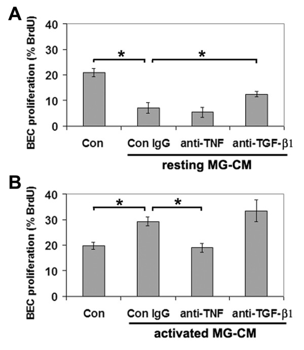
Results: Microglia regulated BEC proliferation in a biphasic manner; microglia conditioned medium (MG-CM) from resting microglia inhibited, while that from activated microglia promoted BEC proliferation. A screen of microglial cytokines revealed that BEC proliferation was inhibited by TGF-β1, but promoted by TNF. ELISA showed that TNF and TGF-β1 were both present in MG-CM, and that while TGF-β1 dominated in resting MG-CM, TNF levels were massively increased in activated MG-CM, shifting the balance in favor of TNF. Antibody-blocking studies revealed that the influence of MG-CM to inhibit or promote BEC proliferation was largely attributable to the cytokines TGF-β1 and TNF, respectively.

Conclusion: This data suggests that microglial activation state might be an important determinant of cerebral angiogenesis; inhibiting BEC proliferation and neovascularization in the normal central nervous system (CNS), but stimulating the growth of new capillaries under neuroinflammatory conditions.


Figures

Figure 1
Figure 1
Regulation of BEC proliferation by microglial-conditioned medium. A. Dual immunocytochemistry for BrdU and Hoechst demonstrated that BEC proliferation was inhibited by conditioned media from resting microglia (MG-CM (rest)), but promoted by conditioned media from activated microglia (MG-CM (activ)). Scale bar = 100 μm. B. Quantification of BEC proliferation in response to microglia-conditioned media (MG-CM) and astrocyte-conditioned media (A-CM). BEC proliferation was examined over 16 hours, and expressed as the percentage of BEC that incorporated BrdU; all points represent the mean ± SEM of 4 experiments. MG-CM from resting or activated microglia, inhibited or promoted BEC proliferation, respectively. A-CM from resting and activated astrocytes both inhibited BEC proliferation. p < 0.05.
Figure 2
Figure 2
Quantification of BEC proliferation in response to cytokines. BEC proliferation was examined over 16 hours in the presence of different concentrations of the cytokines, IFN-α (A), IFN-γ (B), IL-6 (C), TGF-β1 (D), or TNF (E). BEC proliferation is expressed as the percentage of BEC that incorporated BrdU; all points represent the mean ± SEM of 4 experiments. Note that BEC proliferation was significantly stimulated by TNF, and inhibited by TGF-β1. p < 0.05.
Figure 3
Figure 3
ELISA quantification of TNF (A) and TGF-β1 (B) production by microglia or astrocytes, under resting or LPS-activated conditions. All points are expressed in pg/ml concentration of cytokine and represent the mean ± SEM of 4 experiments. Microglial activation resulted in a massive increase in TNF production (50-fold), while astrocytes produced no TNF, even after LPS stimulation. Activation had no significant influence on TGF-β1 production by either cell type.
Figure 4
Figure 4
Function-blocking experiments to examine the role of TNF and TGF-β1 in mediating the effects of MG-CM. BEC proliferation is expressed as the percentage of BEC that incorporated BrdU; all points represent the mean ± SEM of 4 experiments. Note that the inhibitory influence of resting MG-CM was partially relieved by TGF-β1 antibodies (A) and conversely, the mitogenic influence of activated MG-CM was abrogated by anti-TNF antibodies (B). p < 0.05.

References

    1. Marin-Padilla M. Early vascularization of the embryonic cerebral cortex: Golgi and electron microscopic studies. J Comp Neurol. 1985;241:237–249. doi: 10.1002/cne.902410210. - DOI - PubMed
    1. Hayashi T, Noshita N, Sugawara T, Chan PH. Temporal profile of angiogenesis and expression of related genes in the brain after ischemia. J Cereb Blood Flow Metab. 2003;23:166–180. doi: 10.1097/00004647-200302000-00004. - DOI - PubMed
    1. Plate KH, Risau W. Angiogenesis in malignant gliomas. Glia. 1995;15:339–347. doi: 10.1002/glia.440150313. - DOI - PubMed
    1. Desai BS, Schneider JA, Li JL, Carvey PM, Hendey B. Evidence of angiogenic vessels in Alzheimer's disease. J Neural Transm. 2009;116:587–597. doi: 10.1007/s00702-009-0226-9. - DOI - PMC - PubMed
    1. Holley JE, Newcombe J, Whatmore JL, Gutowski NJ. Increased blood vessel density and endothelial cell proliferation in multiple sclerosis cerebral white matter. Neuosci Lett. 2010;470:65–70. doi: 10.1016/j.neulet.2009.12.059. - DOI - PubMed

Publication types