Review
. 2011 Mar;1808(3):818-39.
doi: 10.1016/j.bbamem.2010.11.027. Epub 2010 Dec 4.

An NMR database for simulations of membrane dynamics

Affiliations
Review

An NMR database for simulations of membrane dynamics

Avigdor Leftin et al. Biochim Biophys Acta. 2011 Mar.

Abstract

Computational methods are powerful in capturing the results of experimental studies in terms of force fields that both explain and predict biological structures. Validation of molecular simulations requires comparison with experimental data to test and confirm computational predictions. Here we report a comprehensive database of NMR results for membrane phospholipids with interpretations intended to be accessible by non-NMR specialists. Experimental ¹³C-¹H and ²H NMR segmental order parameters (S(CH) or S(CD)) and spin-lattice (Zeeman) relaxation times (T(1Z)) are summarized in convenient tabular form for various saturated, unsaturated, and biological membrane phospholipids. Segmental order parameters give direct information about bilayer structural properties, including the area per lipid and volumetric hydrocarbon thickness. In addition, relaxation rates provide complementary information about molecular dynamics. Particular attention is paid to the magnetic field dependence (frequency dispersion) of the NMR relaxation rates in terms of various simplified power laws. Model-free reduction of the T(1Z) studies in terms of a power-law formalism shows that the relaxation rates for saturated phosphatidylcholines follow a single frequency-dispersive trend within the MHz regime. We show how analytical models can guide the continued development of atomistic and coarse-grained force fields. Our interpretation suggests that lipid diffusion and collective order fluctuations are implicitly governed by the viscoelastic nature of the liquid-crystalline ensemble. Collective bilayer excitations are emergent over mesoscopic length scales that fall between the molecular and bilayer dimensions, and are important for lipid organization and lipid-protein interactions. Future conceptual advances and theoretical reductions will foster understanding of biomembrane structural dynamics through a synergy of NMR measurements and molecular simulations.


Figures

Fig. 1
Fig. 1
NMR spectroscopy reveals phospholipid membrane dynamics and structure over a range of timescales. The energy landscape of phospholipid mobility is characterized by segmental fluctuations, molecular diffusion, and viscoelastic membrane deformation. Orientational fluctuations correspond to geometry of interaction via Euler angles Ω and by correlation times τc of the motions. (a) Principal axis system of 13C–1H or C–2H bonds fluctuates due to motions of internal segmental frame (I) with respect to the membrane director axis (D). (b) Diffusive phospholipid motions are described by anisotropic reorientation of molecule-fixed frame (M) with respect to the membrane director axis (D). (c) Liquid-crystalline bilayer lends itself to propagation of thermally excited quasi-periodic fluctuations in membrane curvature expressed by motion of the membrane normal (N) relative to membrane director axis (D). The appropriate range of timescales of various complementary biophysical methods is indicated at the bottom of the figure.
Fig. 2
Fig. 2
Solid-state NMR provides residual quadrupolar couplings (RQCs) and residual dipolar couplings (RDCs) that directly correspond to ensemble-averaged molecular structure. (a) The 2H NMR quadrupolar powder-pattern spectrum (light red line) exhibits axial symmetry characteristic of the liquid-crystalline phase for DMPC at 30 °C. Numerically deconvoluted (de-Paked) 2H NMR spectrum (dark line) reveals quadrupolar splittings ΔνQ(i) for individual methylene segments (i) of acyl chains. (b) Isotropic 13C chemical shifts (δ) and pseudostatic recoupled anisotropic powder patterns are obtained under 13C–1H magic-angle spinning (MAS) using the separated local-field (SLF) pulse sequence DROSS [67]. The solid-state 13C–1H lineshapes reveal magnetic dipolar splittings ΔνD(i) for each of the resolved chemically-shifted resonances. The chemical structure of DMPC and spectral assignments are shown.
Fig. 3
Fig. 3
High-resolution solid-state 13C NMR spectroscopy of membrane lipids reveals chemical shifts and nuclear spin-lattice (R1Z) relaxation rates. Isotropic 13C NMR chemical shift (δ) frequency spectrum obtained under magic-angle spinning (MAS) conditions (6 kHz ± 2 Hz, SPINAL-64 1H-decoupling field strength of 50 kHz) for multilamellar DLPC vesicle dispersion at 30 °C. Inset depicts nuclear spin-lattice relaxation rates (R1Z) for (CH2)n carbons (C4–C11) obtained for small unilamellar vesicles (ves) under stationary high-resolution solution NMR conditions versus those for multilamellar dispersions using solid-state MAS techniques. The molecular structure of DLPC and assignments corresponding to the chemical shift assignments in the13C NMR spectra are shown.
Fig. 4
Fig. 4
Structural parameters for liquid-crystalline membranes are derived from 2H NMR spectral data by applying a simple mean-torque model. Properties of membranes include the hydrocarbon thickness DC and cross-sectional area per lipid 〈A〉. (a) Variation of the average cross-sectional areas 〈A〉 are shown for (■) DLPC, (●) DMPC, (◆) DPPC, and (▲) DSPC at temperatures of 10, 30, 50, 65, and 80 °C using data from Ref. [47]. (b) Hydrocarbon thickness DC for homologous saturated phosphatidylcholines shows reduction with increasing temperature for the data set as in (a). Inset depicts the structural parameters of a phospholipid bilayer with number of lipids nL and total volume given by 2nLA〉 (DC + DH), where DH is the headgroup thickness. Phospholipid membrane graphics generated with CHARMM-GUI [98].
Fig. 5
Fig. 5
High-resolution 13C NMR spin lattice relaxation rate profiles for liquid-crystalline DPPC show a significant frequency dispersion characteristic of a motional hierarchy. Spin-lattice relaxation rates R1Z(i) are presented for various carbon positions (i) of DPPC at 50 °C at Larmor frequencies of (formula image) 15.04 MHz, (formula image
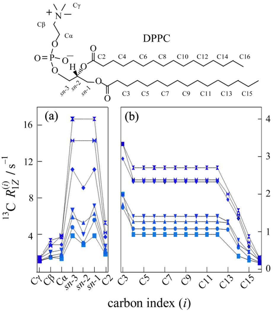
) 20.00 MHz, (◆) 25.15 MHz, (▼) 45.29 MHz, (▲) 90.80 MHz, (●) 125.76 MHz, and (■) 150.84 MHz. (a) The glycerol backbone and choline headgroup are resolved in natural-abundance 13C NMR in the 50–80 ppm region and exhibit a pronounced dispersion of the relaxation rates. (b) Acyl chain segments are observed in the 0–40 ppm fingerprint region of the high-resolution 13C NMR spectrum. The molecular structure of DPPC and assignments corresponding to the carbon index (i) are shown. Data taken in part from Ref. [96].
Fig. 6
Fig. 6
Frequency dispersion of 13C NMR and 2H NMR spin-lattice relaxation rates for liquid-crystalline DMPC bilayers. Nuclear resonance frequencies for 13C or 2H nuclei at a given value of the magnetic field are indicated by ν0 = ω0/2π ≡ νC or νD, respectively. Non-exponential relaxation is evident by comparative fitting of models for intrinsically distributed phospholipid dynamics within the membrane bilayer. (a) 13C NMR relaxation dispersion for (●) (CH2)n carbons (C4–C11) of DMPC (ν0 = 15–150 MHz), and (b) 2H NMR relaxation dispersion for (◆) selectively deuterated C7 carbons of 1,2[7′, 7′-2H] DMPC (ν0 = 3–95 MHz) at 30 °C. By combining the two rate dispersions the frequency range is expanded and a simultaneous best fit is obtained using a composite membrane deformation model (——). Alternatives include molecular diffusion model (– • –), 2D flexible surface model (smectic deformation) (• • •), and (nematic-like) 3D membrane deformation model (– – –). For the composite membrane deformation model, molecular motion is described by principal values D =1.60 ×10⁸ s−1 and D = 2.62 ×10⁶ s−1 of the anisotropic rotational diffusion tensor. Orientational fluctuations of the lipid with respect to the time-averaged membrane normal are described by |Sslow| = 0.89 yielding βMD ≈ 16°. The viscoelastic constant for bilayer dynamics is C = 2.16×10⁻⁶ s−1/2. Data taken in part from Refs. [46,96].
Fig. 7
Fig. 7
Multinuclear NMR spin-lattice relaxation rates for liquid-crystalline bilayers are unified in terms of a frequency power-law. Scaled 2H NMR and 13C NMR spin-lattice relaxation rates 1Z may be compared simultaneously (ν0 ≡ νC, νD). (a) Relaxation rate dispersions for natural-abundance 13C DMPC and isotopically enriched 1,2[3′, 3′-2H] DMPC are shown at 30 °C. The power-law dispersions for the C3 position in (■) 13C NMR and (formula image
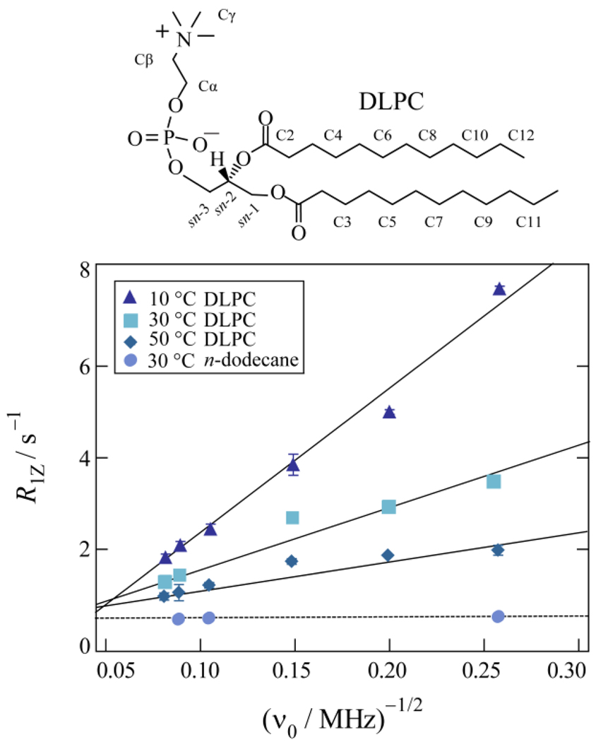
) 2H NMR are fit by a single power-law function (——) with n = −1/2 consistent with a membrane deformation model. (b) Double-logarithmic plots of scaled relaxation rate dispersion with comparative fitting to alternative power-law frequency scalings as indicated. Power-law exponents are shown for n = −2, −1, and −1/2 corresponding to molecular diffusion, flexible surface (smectic deformation), and membrane deformation (nematic-like) models. Data taken in part from Refs. [46,96].
Fig. 8
Fig. 8
Frequency power-law scaling describes non-exponential relaxation of bilayers of phosphatidylcholines over a broad temperature range in the liquid-crystalline state. Model-free analysis for natural-abundance 13C NMR spin-lattice relaxation rate dispersions reveals a single power-law for a homologous series of saturated phosphatidylcholines (ν0 = νC). Frequency scaling is demonstrated for the (CH2)n carbons of (▲) DLPC at 10 °C, (■) DMPC at 30 °C, and (●) DPPC at 50 °C. Each experimental data set exhibits a best fit (– – –) close to the theoretical value of n = −1/2 (––––) showing influence of acyl chain length on membrane dynamics. Data taken in part from Ref. [96].
Fig. 9
Fig. 9
Frequency dispersion of 13C spin-lattice relaxation rates of liquid-crystalline DLPC membrane bilayers compared to liquid hydrocarbon. Relaxation rates obtained for the (CH2)n segments of DLPC are shown at (▲) 10 °C, (■) 30 °C, and (◆) 50 °C. The slope of the power-law relaxation dispersion (ν0 = νC) decreases with an increase in temperature versus corresponding data (●) for (CH2)n carbons of n-dodecane at 30 °C. Extrapolating DPLC relaxation rates to high-frequency or high temperature approximately matches results for n-dodecane in the isotropic liquid state. The center of the phospholipid membrane at high temperature resembles isotropic motion of the hydrocarbon liquid. The molecular structures of n-dodecane and DLPC are shown. Data taken in part from Ref. [96].
Fig. 10
Fig. 10
Natural-abundance 13C NMR spin-lattice relaxation profiles of polyunsaturated lipid bilayers show striking influences from double bonds of acyl chains. Unsaturation in the lipid membranes is observed directly for the polyunsaturated DDPC lipid. Measurements at 50 °C and Larmor frequencies (ν0 = νC) of (▼) 45.29 MHz, (▲) 90.80 MHz, and (●) 125.76 MHz reveal a pronounced variation in relaxation rate. The molecular structure and assignments corresponding to the carbon index (i) are shown. Data taken in part from Ref. [201].

References

    1. Seelig J. Deuterium magnetic resonance: theory and application to lipid membranes. Q. Rev. Biophys. 1977;10:353–418. - PubMed
    1. Griffin RG. Solid state nuclear magnetic resonance of lipid bilayers. Method Enzymol. 1981;72:108–174. - PubMed
    1. Davis JH. The description of membrane lipid conformation, order and dynamics by 2H-NMR. Biochim. Biophys. Acta. 1983;737:117–171. - PubMed
    1. Bloom M, Evans E, Mouritsen OG. Physical properties of the fluid lipid-bilayer component of cell membranes: A perspective. Quart. Rev. Biophys. 1991;24:293–397. - PubMed
    1. Brown MF. Membrane structure and dynamics studied with NMR spectroscopy. In: Merz K Jr, Roux B, editors. Biological Membranes. A Molecular Perspective from Computation and Experiment. Basel: Birkhäuser; 1996. pp. 175–252.

Publication types

LinkOut - more resources