. 2011 Feb 18;406(2):205-14.
doi: 10.1016/j.jmb.2010.11.051. Epub 2010 Dec 4.

HIV-1 Gag extension: conformational changes require simultaneous interaction with membrane and nucleic acid

Affiliations

HIV-1 Gag extension: conformational changes require simultaneous interaction with membrane and nucleic acid

Siddhartha A K Datta et al. J Mol Biol. .

Abstract

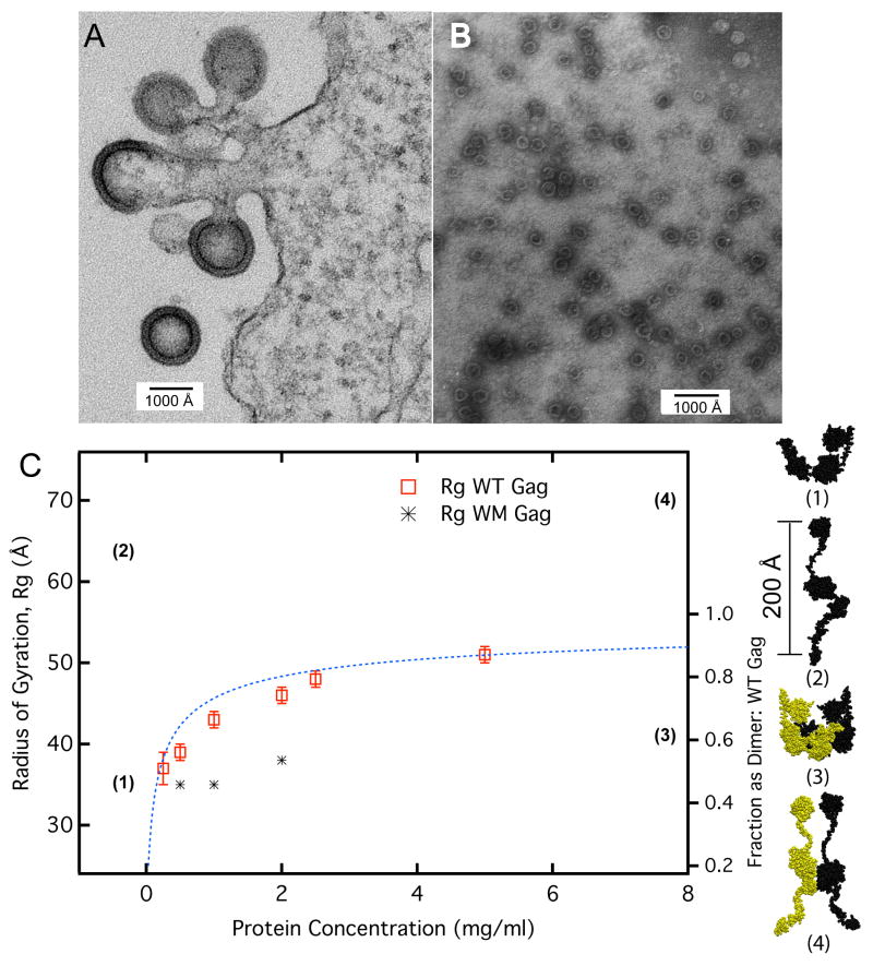
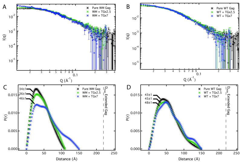
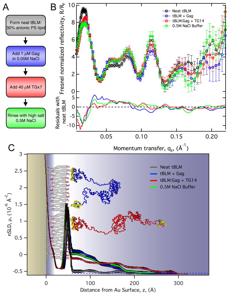
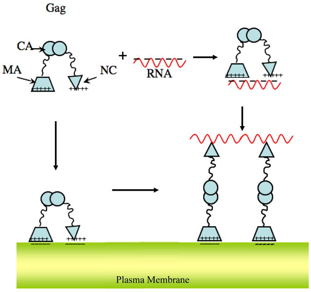
The retroviral Gag polyprotein mediates viral assembly. The Gag protein has been shown to interact with other Gag proteins, with the viral RNA, and with the cell membrane during the assembly process. Intrinsically disordered regions linking ordered domains make characterization of the protein structure difficult. Through small-angle scattering and molecular modeling, we have previously shown that monomeric human immunodeficiency virus type 1 (HIV-1) Gag protein in solution adopts compact conformations. However, cryo-electron microscopic analysis of immature virions shows that in these particles, HIV-1 Gag protein molecules are rod shaped. These differing results imply that large changes in Gag conformation are possible and may be required for viral formation. By recapitulating key interactions in the assembly process and characterizing the Gag protein using neutron scattering, we have identified interactions capable of reversibly extending the Gag protein. In addition, we demonstrate advanced applications of neutron reflectivity in resolving Gag conformations on a membrane. Several kinds of evidence show that basic residues found on the distal N- and C-terminal domains enable both ends of Gag to bind to either membranes or nucleic acid. These results, together with other published observations, suggest that simultaneous interactions of an HIV-1 Gag molecule with all three components (protein, nucleic acid, and membrane) are required for full extension of the protein.


Figures

Figure 1
Figure 1
Gag dimensions. (A) Gag assembly at different stages of immature virion formation imaged by EM as described in supplementary methods. Protein shell in both the spherical virion and incomplete arcs has a thickness of ≈ 200 Å. (B) Recombinant HIV Gag VLPs assembled in vitro with yeast tRNA under standard conditions (described in supplementary methods) at physiological salt buffers and visualized by negative stain EM. These VLPs have a diameter 3× smaller than that of native virions and the protein shell is only 70 to Å 80 Å in thickness. (C) Average protein size in solution as a function of protein concentration. Radius of gyration (Rg) was measured by SANS for WT Gag and WM Gag. The percent of WT Gag molecules as dimer was calculated (dashed blue line) using a Kd of 3.9 μM in D2O. WM Gag has a binding constant 100× weaker than WT and was assumed to be pure monomer over this concentration range. Models for Gag monomer and dimers are shown on the right based on SANS results for the compact state and Cryo EM data for the extended state. The dimerization interface solved by solution NMR for the CA domain was used as a template for generating the dimer complex. The calculated radius of gyrations for both the compact/extended and monomer/dimer configurations are shown on the plot by their numerical designation. (1), monomer in solution; (2), extended monomer, as in authentic immature virions; (3), dimer of (1); (4), dimer of (2).
Figure 2
Figure 2
The influence of single stranded DNA on Gag conformation. WM and WT Gag protein at 20 μM were incubated with short nucleic acid segments containing a TG base sequence repeat. Two different lengths TG×2.5 and TG×7 were used at 240 μM and 80 μM respectively. The SANS spectra of WM Gag and WT Gag are shown in Panel A-B. The pair-distance distribution P(r) determined from the SANS data are given in Panels C & D with Rg values for each condition indicated on the plot. Incubation was performed at 0.5 M NaCl to inhibit protein condensation in the presence of nucleic acid. Gag-TG binding constant, Kd, was determined to be < 1 μM by independent fluorescence anisotropy measurements (see Suppl. Fig. 2). Excess nucleic acid assured complete Gag binding in the SANS experiment.
Figure 3
Figure 3
NR results of WT Gag protein interacting with a tBLM indicates conditions that alter Gag conformation. Panel A, the sequence of measurements performed in situ on the reflectometry instrument. The steps are: 1) Formation of a complete tBLM. 2) Binding WT Gag (buffer: 0.05M NaCl, .001M NaPO4, 5mM TCEP, pH 7.4). 3) Binding of TG×7 DNA to the Gag protein layer. 4) Disassociation of TG×7 using a high ionic strength buffer (same as binding buffer except 0.5 M NaCl). For each experimental condition, three different isotopic aqueous buffer contrasts (mixtures of H2O and D2O) were used. Panel B, the resulting reflectivity spectra for the series of measurements showing the pure H2O buffer data only. Differences in reflectivity from the neat tBLM condition are given as residuals in the bottom of the panel. Panel C, a 1-D nSLD profile of the membrane and Gag determined by fitting the reflectivity data to a “slab model” (see main text for details). Line widths represent the 95 % confidence limits. The inset showing WT Gag cartoons are illustrative models of protein conformations consistent with the overall dimensions determined by reflectivity.
Figure 4
Figure 4
A model for Gag extension consistent with structural characterization and known binding interactions of the protein. The Gag molecule is compact in solution, even when bound to RNA. RNA binds to the NC domain with very high affinity, but evidently can also bind, with lower affinity, to the MA domain. The MA domain targets and anchors Gag to the anionic surface of the plasma membrane. Additionally the NC domain may also associate with the membrane through electrostatic interactions. Only in the presence of all three components i) protein, ii) nucleic acid and iii) membrane, extended Gag is formed. A plausible mechanism is one in which the viral RNA and the membrane separate the terminal domains through their preferential interaction. Cross-linking several membrane bound Gag molecules together by the RNA strand and inter-protein interactions may further stabilize extended Gag.

References

    1. Swanstrom R, Wills JW. Synthesis, assembly, and processing of viral proteins. In: Coffin JM, Hughes SH, Varmus HE, editors. Retroviruses. Cold Spring Harbor Laboratory Press; Plainview: 1997. pp. 263–334. - PubMed
    1. Ganser-Pornillos BK, Yeager M, Sundquist WI. The structural biology of HIV assembly. Curr Opin Struct Biol. 2008;18:203–217. - PMC - PubMed
    1. Zhou WJ, Parent LJ, Wills JW, Resh MD. Identification of a membrane binding domain within the amino-terminal region of human immunodeficiency virus type 1 Gag protein which interacts with acidic phospholipids. J Virol. 1994;68:2556–2569. - PMC - PubMed
    1. Fuller SD, Wilk T, Gowen BE, Kräusslich HG, Vogt VM. Cryo-electron microscopy reveals ordered domains in the immature HIV-1 particle. Curr Biol. 1997;7:729–738. - PubMed
    1. Datta SAK, Curtis J, Ratcliff W, Clark P, Crist R, Lebowitz J, Krueger S, Rein A. Conformation of the HIV-1 Gag Protein in Solution. J Mol Biol. 2007;365:812–824. - PMC - PubMed

Publication types

LinkOut - more resources