. 2011 Apr;174(1):203-12.
doi: 10.1016/j.jsb.2010.11.027. Epub 2010 Dec 4.

Self-aligning amelogenin nanoribbons in oil-water system

Affiliations

Self-aligning amelogenin nanoribbons in oil-water system

Xiaodong He et al. J Struct Biol. 2011 Apr.

Abstract

The highly organized microstructure of dental enamel is a result of protein-guided anisotropic growth of apatite nanofibers. It is established that amelogenin proteins, the main constituent of the developing enamel matrix, form nanospheres in vitro, but the amphiphilic nature of the full-length protein conveys the possibility of generating more complex structures as observed with other surfactant-like molecules. This study tested if the use of metastable oil-water emulsions can induce supramolecular assemblies of amelogenin. Recombinant full-length amelogenin, rH174, was mixed into octanol/ethyl acetate preparations of different ratios to form emulsions at pH 4.5 and 7.4. Atomic force and electron microscopy showed the formation of 16.7±1.0nm wide nanoribbons which grew to several micrometer length over a period of days. Nanoribbons formed from reverse micelles by enabling hydrophobic tails of the molecules to interact while preventing the formation of amelogenin nanospheres. Ribbon formation required the presence of calcium and phosphate ions and may be localized at a dark central line along the amelogenin ribbons. The ribbons have a strong tendency to align in parallel maintaining 5-20nm space between each other. The growth rates and number of ribbons were significantly higher at pH 4.5 and related to the metastability of the emulsion. A model for ribbon extension proposes the addition of short segments or amelogenin dimers to the ends of the ribbon. The formation of self-aligning and uniaxially elongating amelogenin structures triggered by the presence of calcium and phosphate may represent a suitable new model for protein controlled mineralization in enamel.


Figures

Figure 1
Figure 1
Emulsions of aqueous amelogenin suspensions form when mixed with Octanol/Ethylacetate in the presence of calcium and phosphate, t = 10 min (a); emulsions prepared at pH 4.5 gradually phase separated into an oil and water phase, t = 3h (b) and are completely separated within 48 hours (c). Particle size analysis by time-resolved dynamic light scattering (d) (initial 48 hours not shown due to particle size being out of range) shows a decrease after 2 days from micrometer sized particles to sizes of 30 – 100 nm followed by fluctuations in particle sizes with particles reaching several micrometers in diameter at 6–7 days of incubation at 37°C.
Figure 2
Figure 2
TEM-images of amelogenin rH174 at different times of incubation; top row (a–c) rH174 was dissolved in aqueous suspensions at pH 2–3, subsequently pH was raised 4.5 using KOH and Tris-buffer. The characteristic 20 nm nanosphere of amelogenin was observed at all time points between 1 to 14 days of incubation, t = 24 h (a); occasionally nanosphere aggregated into short strings, t = 48 h (b), and t = 7 days (c). When aqueous amelogenin suspensions were mixed at pH 2–3 with a non-polar solvent (octanol/ethyl acetate) and subsequently pH was raised to 4.5, ribbon-like nano-structures appeared as early as 24 hours (d). Ribbons grew in length over time and arranged themselves in a parallel manner, t = 7 days (e); ribbons reached several micrometer in length and formed well aligned bundles of nanoribbons, t= 14 days (f).
Figure 3
Figure 3
TEM analysis of self-assembled amelogenin supramolecular structure showed that structures are about 17 nm wide and are thin ribbons, only 3 nm thick. The structures tend to align themselves in a parallel fashion, most likely through electrostatic repulsion at the long axes of the ribbons.
Figure 4
Figure 4
TEM analysis of amelogenin nanoribbons showed the presence of a dark central line which was present in the absence of stain and when specimens were positively or negatively stained with nano-W (a). Averaging over 604 equally sized ribbon segments (negative stain) further revealed the evidence of a dark central line that indicated higher electron density which could be associated with the presence of calcium and/or phosphate ions which were both required for self-assembly into ribbons.
Figure 5
Figure 5
AFM topographic images (tapping mode) of a series of amelogenin nanostructures that formed at pH 4.5 (a–c) or pH 7.4 (d–f). Ribbons were observed as early as 30 min after emulsion formation at pH 4.5 (a), while ribbons appeared after 3 days at pH 7.4 (d). Ribbons became more abundant with time at pH 4.5, at 7 days (b) and at 14 days (c) while ribbons remained scarce at pH 7.4.
Figure 6
Figure 6
Large amounts of amelogenin ribbons were observed by SEM (a) and elemental analysis by EDX confirmed the presence of small amounts of Calcium and Phosphate ions in these structures which were deposited onto a silica based microscope slide (b). The more randomly distributed ribbons were also observed by AFM but only in areas were large amounts of the protein could be immobilized to the glass surface (c). Analysis of the fibrillar structures using micro-Raman spectroscopy showed large similarities between the lyophilized amelogenin powder as received after purification and the self-assembled structures deposited onto a glass-slide. Overall the spectrum obtained from the amelogenin ribbons appeared more pronounced with respect to the amide vibrational modes above 1400 cm−1. In particular the sharpening of a band at 1670 cm−1 and the occurrence of a band around 1610 cm−1 are indicative of the formation of β-sheet structures.
Figure 7
Figure 7
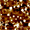
AFM image of micellar structures that are present at the early time points of emulsification. Sample obtained at 5 min after vortexing.
Figure 8
Figure 8
Schematic drawing of the possible supramolecular structure of amelogenin nanoribbons that form at the oil-water interface. Initially a reverse micelle forms with the hydrophobic portion of the molecules (red) exposed to the surface. The hydrophobic portions can now interact with each other and form unique bonds that are most likely based on calcium bridges and Van der Waals bonds and lead to the formation of structural motifs like β-sheets. Such amelogenin dimers will grow by the addition of other dimers that are released from the reverse micelles soon as these micelles disintegrate at the water-oil interface. Gradually ribbons will form by addition of segments to its ends. Segments repulse each other at the long axis to electrostatic charges that derive from the hydrophilic portion (blue) lining the long axis of the ribbons. Electrostatic repulsion will result in parallel alignment of ribbons over time and also facilitates oriented grows of ribbons along their long axes.

Similar articles

Cited by

References

    1. Aichmayer B, Margolis HC, Sigel R, Yamakoshi Y, Simmer JP, Fratzl P. The onset of amelogenin nanosphere aggregation studied by small-angle X-ray scattering and dynamic light scattering. J Struct Biol. 2005;151:239–49. - PubMed
    1. Aichmayer B, Wiedemann-Bidlack FB, Gilow C, Simmer JP, Yamakoshi Y, Emmerling F, Margolis HC, Fratzl P. Amelogenin nanoparticles in suspension: Deviations from spherical shape and pH-dependent aggregation. Biomacromolecules. 2010;11:369–76. - PMC - PubMed
    1. Beniash E, Simmer JP, Margolis HC. The effect of recombinant mouse amelogenins on the formation and organization of hydroxyapatite crystals in vitro. J Struct Biol. 2005;149:182–90. - PubMed
    1. Berne BJ, Pecora R. Laser Light-Scattering from Liquids. Annual Review of Physical Chemistry. 1974;25:233–253.
    1. Chen Y, Wu QS, Dingibi Y. Stepwise assembly of nanoparticies, -tubes, -rods, and -wires in reverse micelle systems. European Journal of Inorganic Chemistry. 2007:4906–4910.

Publication types

LinkOut - more resources