

Food allergy herbal formula 2 protection against peanut anaphylactic reaction is via inhibition of mast cells and basophils
- PMID: 21134573
- PMCID: PMC3059770
- DOI: 10.1016/j.jaci.2010.09.013
Food allergy herbal formula 2 protection against peanut anaphylactic reaction is via inhibition of mast cells and basophils
Abstract
Background: Mast cells and basophils are key effector cells of IgE-mediated anaphylactic reactions. The Chinese herbal formula, food allergy herbal formula 2 (FAHF-2), protects against peanut anaphylaxis in mice. However, the mechanisms underlying this effect are not fully elucidated.
Objective: To investigate whether FAHF-2 inhibits mast cell/basophil numbers and IgE-mediated activation.
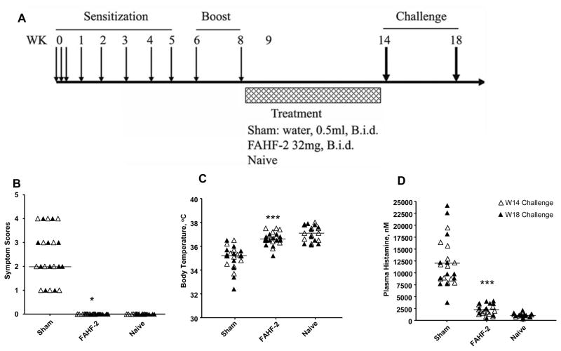
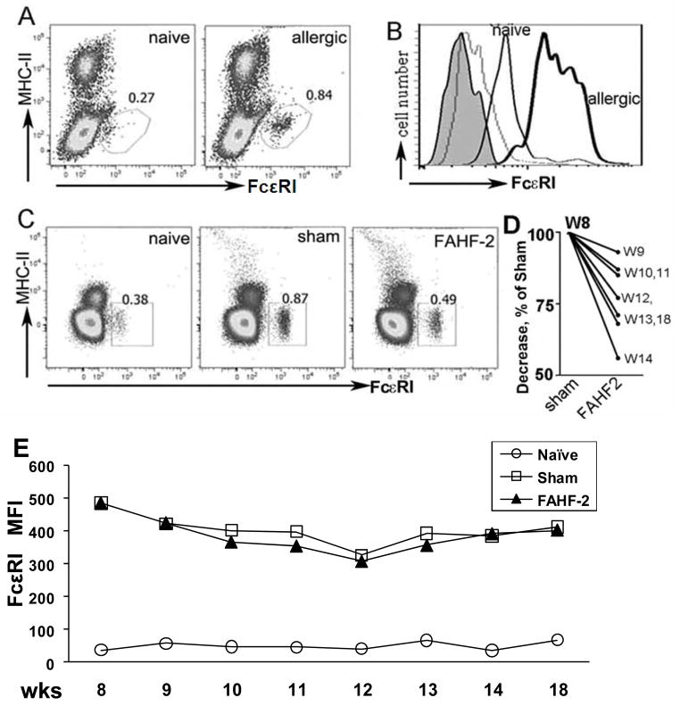
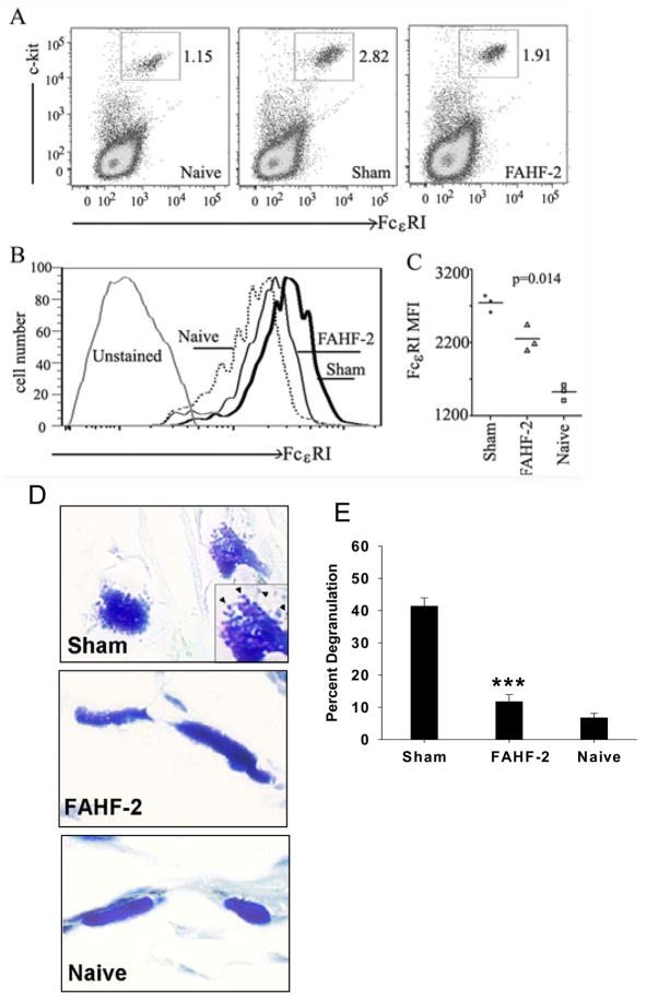
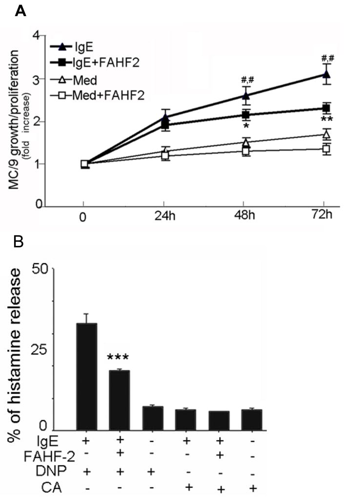
Methods: Mice with peanut allergy (PNA mice) were treated with FAHF-2 intragastrically for 7 weeks and challenged intragastrically with peanut 1 day and 4 weeks posttreatment. Peripheral blood basophil numbers and peritoneal mast cell numbers and FcεRI expression were determined. Direct effects of FAHF-2 on the murine mast cell line MC/9, and effects of 4 fractions and 3 compounds isolated from FAHF-2 on rat basophilic leukemia cells (RBL-2H3) and human skin mast cells degranulation and on the IgE-mediated spleen tyrosine kinase signaling pathway, were determined.
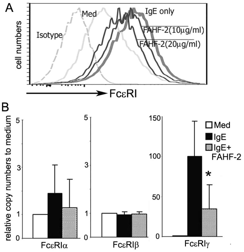
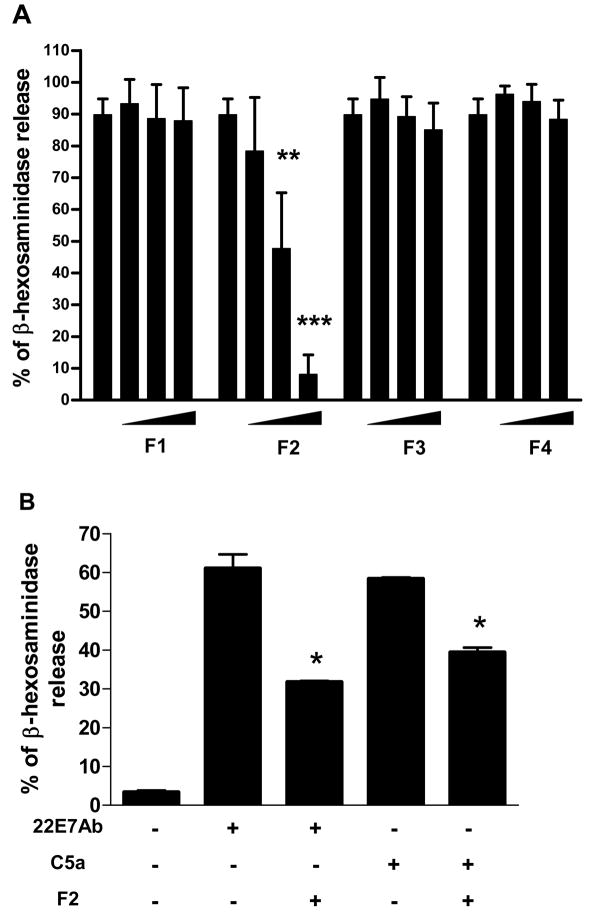
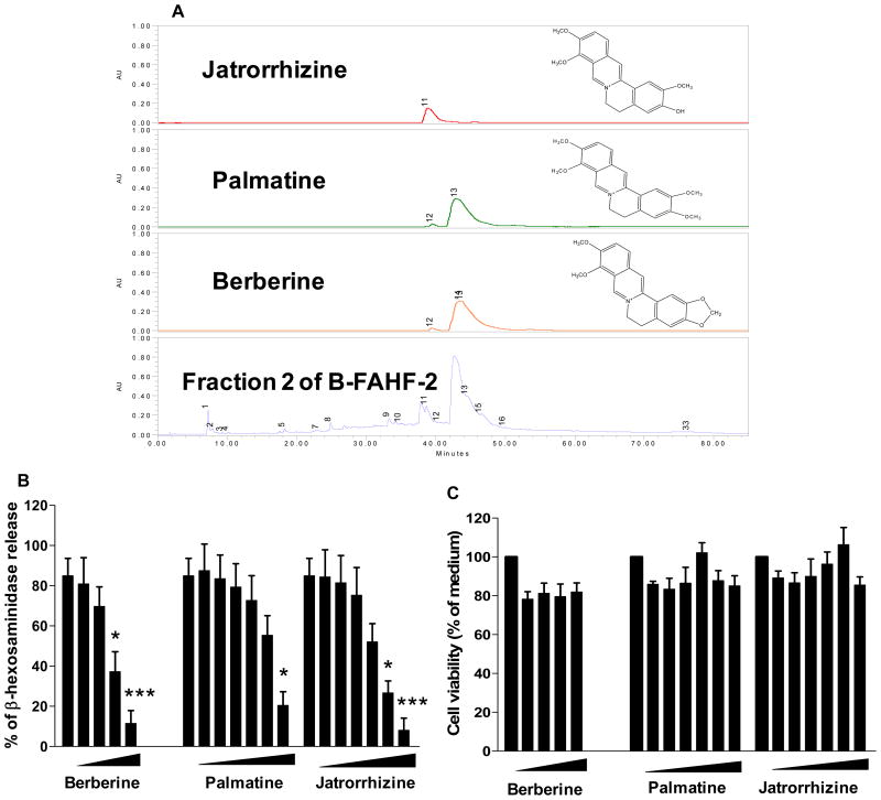
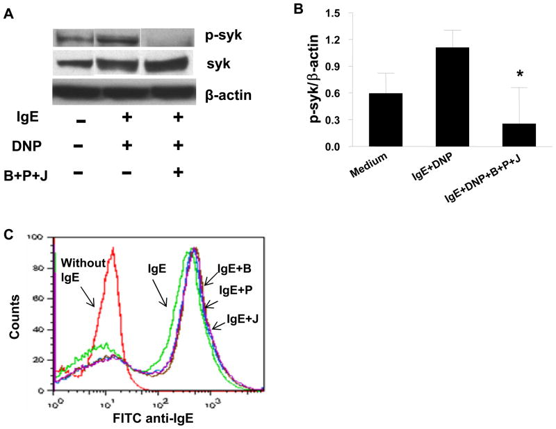
Results: Although all sham-treated PNA mice developed anaphylaxis, FAHF-2-treated PNA mice were protected against anaphylaxis after peanut challenge at 1 day and 4 weeks posttherapy. Reduction of peripheral blood basophils began after 1 week of treatment and continued for at least 4 weeks posttherapy. The number and FcεRI expression of peritoneal mast cells were also significantly decreased 4 weeks posttherapy. FAHF-2-treated MC/9 cells showed significantly reduced IgE-induced FcεRI expression, FcεRI γ mRNA subunit expression, proliferation, and histamine release on challenge. Fraction 2 from FAHF-2 inhibited RBL-2H3 cell and human mast cell degranulation. Three compounds from fraction 2-berberine, palmatine, and jatrorrhizine-inhibited RBL-2H3 cell degranulation via suppressing spleen tyrosine kinase phosphorylation.
Conclusion: Food allergy herbal formula 2 reduction of basophils and mast cell numbers as well as suppression of IgE-mediated mast cell activation may contribute to FAHF-2's persistent protection against peanut anaphylaxis.
Copyright © 2010 American Academy of Allergy, Asthma & Immunology. Published by Mosby, Inc. All rights reserved.
Conflict of interest statement
Figures
Similar articles
-
Food Allergy Herbal Formula-1 (FAHF-1) blocks peanut-induced anaphylaxis in a murine model.J Allergy Clin Immunol. 2001 Oct;108(4):639-46. doi: 10.1067/mai.2001.118787. J Allergy Clin Immunol. 2001. PMID: 11590394
-
The Chinese herbal medicine formula FAHF-2 completely blocks anaphylactic reactions in a murine model of peanut allergy.J Allergy Clin Immunol. 2005 Jan;115(1):171-8. doi: 10.1016/j.jaci.2004.10.003. J Allergy Clin Immunol. 2005. PMID: 15637565
-
Efficacy, safety and immunological actions of butanol-extracted Food Allergy Herbal Formula-2 on peanut anaphylaxis.Clin Exp Allergy. 2011 Apr;41(4):582-91. doi: 10.1111/j.1365-2222.2010.03643.x. Epub 2010 Dec 1. Clin Exp Allergy. 2011. PMID: 21121976 Free PMC article.
-
Targeting the FcεRI Pathway as a Potential Strategy to Prevent Food-Induced Anaphylaxis.Front Immunol. 2020 Dec 17;11:614402. doi: 10.3389/fimmu.2020.614402. eCollection 2020. Front Immunol. 2020. PMID: 33391286 Free PMC article. Review.
-
IgE and IgG Antibodies as Regulators of Mast Cell and Basophil Functions in Food Allergy.Front Immunol. 2020 Dec 11;11:603050. doi: 10.3389/fimmu.2020.603050. eCollection 2020. Front Immunol. 2020. PMID: 33362785 Free PMC article. Review.
Cited by
-
Mouse Models for Food Allergies: Where Do We Stand?Cells. 2019 Jun 6;8(6):546. doi: 10.3390/cells8060546. Cells. 2019. PMID: 31174293 Free PMC article. Review.
-
Oral and sublingual immunotherapy for food allergy: current progress and future directions.Curr Opin Immunol. 2013 Dec;25(6):781-7. doi: 10.1016/j.coi.2013.07.011. Epub 2013 Aug 20. Curr Opin Immunol. 2013. PMID: 23972904 Free PMC article. Review.
-
Future Therapies for IgE-Mediated Food Allergy.Curr Pediatr Rep. 2014 Jun 1;2(2):119-126. doi: 10.1007/s40124-014-0041-0. Curr Pediatr Rep. 2014. PMID: 24883237 Free PMC article.
-
The Future of Food Allergy Management: Advancements in Therapies.Curr Allergy Asthma Rep. 2024 Apr;24(4):161-171. doi: 10.1007/s11882-024-01133-1. Epub 2024 Feb 23. Curr Allergy Asthma Rep. 2024. PMID: 38393624 Review.
-
Inhibition of pathologic immunoglobulin E in food allergy by EBF-2 and active compound berberine associated with immunometabolism regulation.Front Immunol. 2023 Feb 7;14:1081121. doi: 10.3389/fimmu.2023.1081121. eCollection 2023. Front Immunol. 2023. PMID: 36825019 Free PMC article.
References
-
- Wang J, Sampson HA. Food anaphylaxis. Clin Exp Allergy. 2007;37(5):651–60. - PubMed
-
- Galli SJ. Mast cells and basophils. Curr Opin Hematol. 2000;7(1):32–9. - PubMed
-
- Kawakami T, Galli SJ. Regulation of mast-cell and basophil function and survival by IgE. Nat Rev Immunol. 2002;2(10):773–86. - PubMed
-
- Qu C, Srivastava K, Ko J, Zhang TF, Sampson HA, Li XM. Induction of tolerance after establishment of peanut allergy by the food allergy herbal formula-2 is associated with up-regulation of interferon-gamma. Clin Exp Allergy. 2007;37(6):846–55. - PubMed
-
- Srivastava KD, Kattan JD, Zou ZM, Li JH, Zhang L, Wallenstein S, et al. The Chinese herbal medicine formula FAHF-2 completely blocks anaphylactic reactions in a murine model of peanut allergy. J Allergy Clin Immunol. 2005;115(1):171–8. - PubMed
Publication types
MeSH terms
Substances
Grants and funding
LinkOut - more resources
Full Text Sources
Other Literature Sources
Medical
