. 2010 Dec 8;18(12):1596-607.
doi: 10.1016/j.str.2010.09.020.

Nanometer propagation of millisecond motions in V-type allostery

Affiliations

Nanometer propagation of millisecond motions in V-type allostery

James M Lipchock et al. Structure. .

Abstract

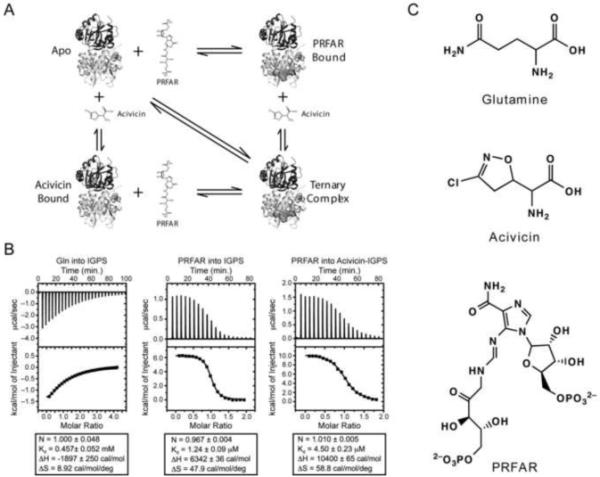
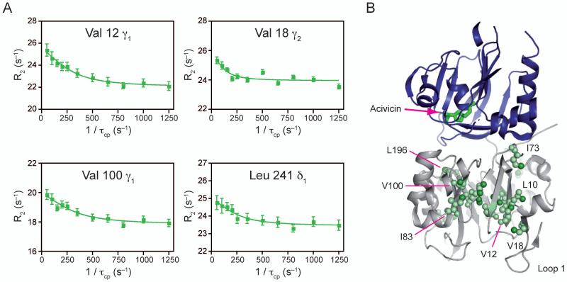
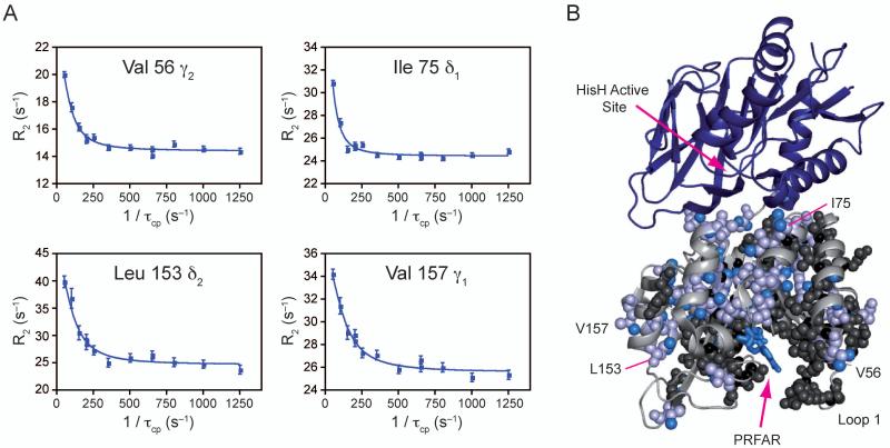
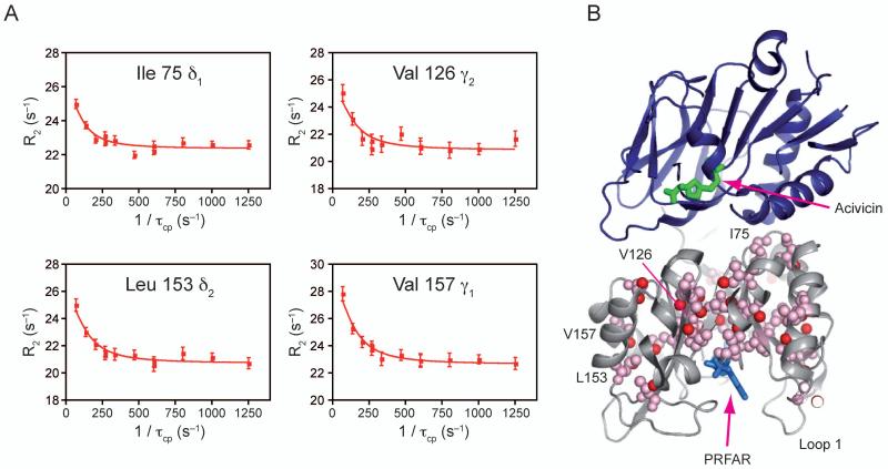
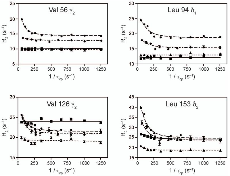
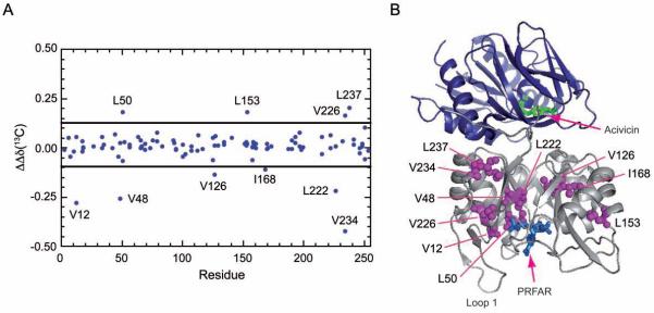
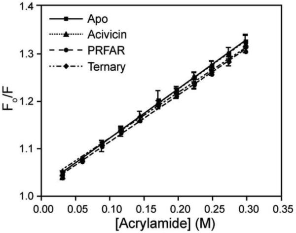
Imidazole glycerol phosphate synthase (IGPS) is a V-type allosteric enzyme, which is catalytically inactive for glutamine hydrolysis until the allosteric effector, N'-[(5'-phosphoribulosyl)formimino]-5-aminoimidazole-4-carboxamide-ribonucleotide (PRFAR) binds 30 Å away. In the apo state, NMR relaxation dispersion experiments indicate the absence of millisecond (ms) timescale motions. Binding of the PRFAR to form the active ternary complex is endothermic with a large positive entropy change. In addition, there is a protein wide enhancement of conformational motions in the ternary complex, which connect the two active sites. NMR chemical shift changes and acrylamide quenching experiments suggest that little in the way of structural changes accompany these motions. The data indicate that enzyme activation in the ternary complex is primarily due to an enhancement of ms motions that allows formation of a population of enzymatically active conformers.


Figures

Figure 1
Figure 1. Ligand interaction with IGPS
A) Thermodynamic cycle of ligand binding to IGPS. The glutamine analog, acivicin, and PRFAR are shown as sticks when unbound and spheres when bound. B) ITC profiles of ligand binding to IGPS. Thermodynamic parameters are shown below each titration C) Structures of glutamine, acivicin and PRFAR.
Figure 2
Figure 2. NMR characterization of μs-ms motions in apo ILV 13CH3 methyl labeled HisF
A) Four representative 13C1H MQ dispersion curves out of the 17 with positive relaxation dispersion amplitudes. B) Structural mapping of ILV dispersion onto IGPS. Residues exhibiting dispersion are shown in light orange spheres. Individual atoms with dispersion are highlighted with bright orange spheres. See also Supplemental Table 1 and Supplemental Figure 1.
Figure 3
Figure 3. NMR characterization of μs-ms motions in acivicin bound ILV 13CH3 methyl labeled HisF
A) Four representative 13C1H MQ dispersion curves out of the 17 with positive relaxation dispersion amplitudes. B) Structural mapping of ILV dispersion onto IGPS. Acivicin is shown bound to HisH in green sticks. Residues exhibiting dispersion are shown in light green spheres. Individual atoms with dispersion are highlighted with bright green spheres. See also Supplemental Table 2.
Figure 4
Figure 4. NMR characterization of μs-ms motions in PRFAR bound 15N and ILV 13CH3 methyl labeled HisF
A) Four representative 13C1H MQ dispersion curves out of the 68 (includes 15N SQ and 13C1H MQ) with positive relaxation dispersion amplitudes. B) Structural mapping of ILV dispersion onto IGPS. PRFAR is shown bound to HisF in blue sticks. Residues exhibiting dispersion are shown in light blue spheres. Individual atoms with dispersion are highlighted with bright blue spheres. Residues undergoing exchange broadening are shown in black spheres. See also Supplemental Table 3.
Figure 5
Figure 5. NMR characterization of μs-ms motions in ternary ILV 13CH3 methyl labeled HisF
A) Four representative 13C1H MQ dispersion curves out of the 38 with positive relaxation dispersion amplitudes. B) Structural mapping of ILV dispersion onto IGPS. PRFAR is shown bound to HisF in blue sticks, while acivicin is shown bound to HisH in green sticks. Residues exhibiting dispersion are shown in pink spheres. Individual atoms with dispersion are highlighted with red spheres. See also Supplemental Table 4.
Figure 6
Figure 6. Comparison of dispersion profiles for the apo, acivicin bound, PRFAR bound and ternary states of ILV 13CH3 methyl labeled HisF
Dispersion overlays highlighting differences in dispersion amplitudes for the four states. Apo data are shown as squares, acivicin bound data are as triangles, PRFAR bound (circles) and ternary complex data as diamonds.
Figure 7
Figure 7. Summary of ms motions
A) Venn-type diagram illustrating the relation between flexible residues from ILV relaxation dispersion experiments and the enzyme complex in which they occur for apo (black), acivicin (green), PRFAR (blue), and ternary (red). Residues common to all four are shown in a larger font size. B) Residues exhibiting ILV methyl 13C1H MQ dispersion in the PRFAR bound and ternary states are shown mapped onto IGPS. Residues with dispersion in only the PRFAR bound state are shown in blue spheres, while those with dispersion in only the ternary complex are shown in red. Residues with dispersion in both states are shown in magenta.
Figure 8
Figure 8. Synergistic chemical shift changes due to ligand binding
The 13C ΔΔδ chemical shift differences between the two singly ligated forms of IGPS and the ternary complex is shown versus residue for the Ile, Leu, and Val positions as calculated with equation (1). Residues with a non-additive ΔΔδ value ≥ 1.5 σ from the mean as indicated by the horizontal black lines are mapped onto the IGPS structure in magenta spheres. Acivicin and PRFAR are shown in green and blue sticks, respectively. Not shown are synergistic 1H chemical shift changes, which show similar behavior. See also Supplemental Figures 2 and 3.
Figure 9
Figure 9. Motions at the HisH active site
HisH is shown in blue cartoon ribbon. The loop comprising the oxyanion hole is highlighted in cyan. The catalytic Cys84 is shown in sticks with the nucleophilic sulfur atom in yellow. The carbonyl oxygen of Gly50 is shown as a red sphere, while the amide proton of Val51 is represented as a gray sphere to emphasize the geometry of the inactive glutamine amidotransferase. Activation of HisH is predicted to require flipping of the amide bond to present the amide proton of Val51 to allow for stabilization of the oxyanion intermediate. B) Resonance broadening of Gly50 in 15N HisH-IGPS upon titration with PRFAR. All spectra were collected under identical conditions with an equal number of scans and are shown contoured equally. Gly50 goes from an intense, well-resolved resonance in the apo form (red) to broadened beyond detection at 0.87 mM PRFAR (purple) (99.8% saturated), with intermediate titration points of 0.038 mM (orange), 0.150 mM (yellow), 0.220 mM (green), and 0.290 mM (blue).
Figure 10
Figure 10. Acrylamide accessibility assay
Comparison of Stern-Volmer tryptophan fluorescence quenching with acrylamide for apo (squares), acivicin bound (triangles), PRFAR bound (circles) and ternary (diamonds) IGPS. For each state, 10 μM IGPS was utilized in 50 mM HEPES pH 8.0 at room temperature.

References

    1. Ackers GK, Doyle ML, Myers D, Daugherty MA. Molecular code for cooperativity in hemoglobin. Science. 1991;255:54–63. - PubMed
    1. Amaro RE, Myers RS, Davisson VJ, Luthey-Schulten ZA. Structural elements in IGP synthase exclude water to optimize ammonia transfer. Biophys J. 2005;89:475–487. - PMC - PubMed
    1. Amaro RE, Sethi A, Myers RS, Davisson VJ, Luthey-Schulten ZA. A network of conserved interactions regulates the allosteric signal in a glutamine amidotransferase. Biochemistry. 2007;46:2156–2173. - PubMed
    1. Beismann-Driemeyer S, Sterner R. Imidazole glycerol phosphate synthase from Thermotoga maritima - Quaternary structure, steady-state kinetics, and reaction mechanism of the bienzyme complex. Journal of Biological Chemistry. 2001;276:20387–20396. - PubMed
    1. Bertrand D, Gopalakrishnan M. Allosteric modulation of nicotinic acetylcholine receptors. Biochem Pharmacol. 2007;74:1155–1163. - PubMed

Publication types

MeSH terms

Substances