. 2011 Feb;79(2):724-31.
doi: 10.1128/IAI.00980-10. Epub 2010 Dec 6.

Native properdin binds to Chlamydia pneumoniae and promotes complement activation

Affiliations

Native properdin binds to Chlamydia pneumoniae and promotes complement activation

Claudio Cortes et al. Infect Immun. 2011 Feb.

Abstract

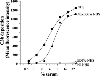
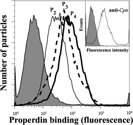
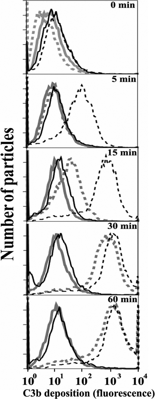
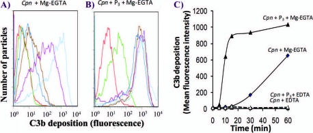
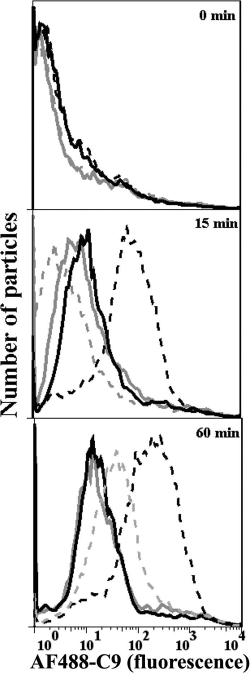
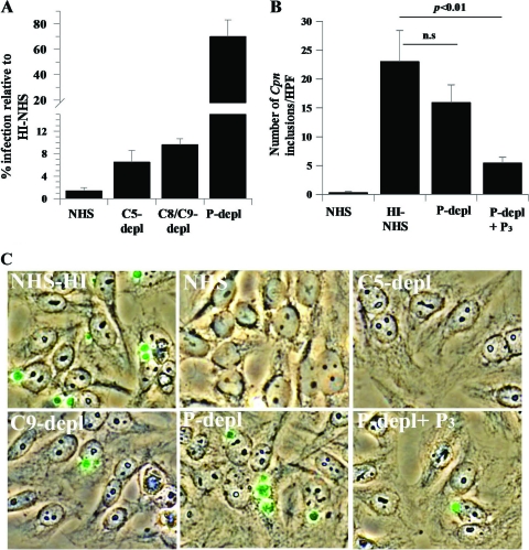
Activation of complement represents one means of natural resistance to infection from a wide variety of potential pathogens. Recently, properdin, a positive regulator of the alternative pathway of complement, has been shown to bind to surfaces and promote complement activation. Here we studied whether properdin-mediated complement activation occurs on the surface of Chlamydia pneumoniae, an obligate intracellular Gram-negative bacterium that causes 10 to 20% of community-acquired pneumonia. We have determined for the first time that the physiological P₂, P₃, and P₄ forms of human properdin bind to the surface of Chlamydia pneumoniae directly. The binding of these physiological forms accelerates complement activation on the Chlamydia pneumoniae surface, as measured by C3b and C9 deposition. Finally, properdin-depleted serum could not control Chlamydia pneumoniae infection of HEp-2 cells compared with normal human serum. However, after addition of native properdin, the properdin-depleted serum recovered the ability to control the infection. Altogether, our data suggest that properdin is a pattern recognition molecule that plays a role in resistance to Chlamydia infection.


Figures

FIG. 1.
FIG. 1.
Activation of the human complement system by Chlamydia pneumoniae. C. pneumoniae bacteria (25 × 106 IFU) were incubated with the indicated concentrations of normal human serum (NHS) in the presence of Ca2+ and Mg2+ to evaluate all complement pathways, with Mg-EGTA to evaluate the alternative pathway, or with EDTA or heat-inactivated (HI) serum (56°C at 30 min) to inhibit all complement system pathways. C. pneumoniae bacteria were incubated for 1 h at 37°C, and complement activation was stopped by adding cold HBSS-EDTA. C. pneumoniae bacteria were then washed, and C3b deposition was assessed by flow cytometry using a PE-labeled anti-C3b antibody. EB-positive cells were gated using an FITC-labeled anti-C. pneumoniae antibody. The mean fluorescence intensity of C3b deposition versus the percentage of serum was plotted. No C3b deposition was detected in the presence of EDTA or when HI-NHS was used.
FIG. 2.
FIG. 2.
Native P2, P3, and P4, forms of properdin bind to Chlamydia pneumoniae. C. pneumoniae bacteria (50 × 106 IFU) were incubated with 3 μg of each form of properdin (purified by ion-exchange chromatography, followed by size exclusion chromatography) in 100 μl HBSS plus 0.1% BSA for 30 min at 4°C. C. pneumoniae bacteria were then washed and analyzed by FACS analysis using an anti-properdin monoclonal antibody or an IgG1 monoclonal antibody isotype control (shaded), followed by an FITC-conjugated anti-mouse IgG antibody. (Inset) The purity of the C. pneumoniae elementary body preparation was assayed by FACS analysis using a C. pneumoniae-specific monoclonal antibody or an IgG2a monoclonal antibody isotype control, followed by an FITC-conjugated anti-mouse IgG antibody. The number of particles versus properdin binding (fluorescence intensity) is shown. Cpn, C. pneumoniae.
FIG. 3.
FIG. 3.
A physiological form of properdin (P3), when bound to C. pneumoniae, induces complement activation using properdin-depleted serum. C. pneumoniae bacteria (50 × 106 IFU) were incubated with (black lines) or without (gray lines) 3 μg of P3 in 100 μl HBSS-0.1% BSA for 30 min at 4°C. C. pneumoniae bacteria were then washed, and 15% properdin-depleted serum was added in the presence of HBSS-0.1% BSA-Mg-EGTA (dashed lines) or HBSS-0.1% BSA-EDTA (solid lines) and incubated at 37°C for the indicated times. The particles were then washed with cold HBSS-EDTA, and C3b deposition was assessed by FACS analysis using an FITC-labeled anti-C3b antibody. The number of particles versus C3b deposition (fluorescence intensity) is shown.
FIG. 4.
FIG. 4.
The physiological form of properdin (P3), when bound to C. pneumoniae, induces complement activation using normal human serum. The P3 form of properdin was isolated from purified properdin by ion-exchange chromatography, followed by size exclusion chromatography, as described in Materials and Methods. (A and B) C. pneumoniae bacteria (50 × 106 IFU) were incubated with (Cpn + P3) or without (Cpn) 3 μg P3 in 100 μl HBSS-0.1% BSA for 30 min at 4°C. C. pneumoniae bacteria were then washed, and 5% normal human serum was added in the presence of HBSS-0.1% BSA-Mg-EGTA or HBSS-0.1% BSA-EDTA. These mixtures were incubated for the indicated times at 37°C. The particles were then washed with cold HBSS plus EDTA, and C3b deposition was assessed by flow cytometry using an FITC-labeled anti-C3 antibody. Red, 0 min; green, 5 min; dark blue, 10 min; brown, 15 min; purple, 30 min; light blue, 60 min. (C) C3b deposition was graphed as mean fluorescence intensity, obtained from panels A and B, versus time.
FIG. 5.
FIG. 5.
The physiological form of properdin (P3) bound to Chlamydia pneumoniae accelerates C9 deposition on EBs. C. pneumoniae bacteria (50 × 106 IFU) were incubated with (black lines) or without (gray lines) 3 μg P3 in 100 μl HBSS-0.1% BSA for 30 min at 4°C. The particles were then washed, and 15% properdin-depleted serum plus Alexa Fluor 488-labeled C9 (AF488-C9) was added in the presence of HBSS-0.1% BSA-Mg-EGTA (dashed lines) or HBSS-0.1% BSA-EDTA (solid lines) for 0, 15, and 60 min at 37°C. The particles were then washed with cold HBSS-EDTA, and Alexa Fluor 488-labeled C9 deposition was assessed by flow cytometry. The number of particles versus C9 deposition (fluorescence intensity) is shown.
FIG. 6.
FIG. 6.
Early complement components and the presence of properdin in serum are important in controlling C. pneumoniae infection. (A) EBs were preincubated with 30% NHS or serum depleted of C5, C8/C9, or properdin for 1 h at 37°C. EBs were then used to infect HEp-2 monolayers as described in Materials and Methods, and the percentage of infection of HEp2 cells was measured relative to that with heat-inactivated serum (not shown). (B) EBs were preincubated with 30% NHS, HI-NHS, or properdin-depleted serum (P-depl), with or without P3 (15 μg/ml), in RPMI 1640 plus Ca2+/Mg2+ for 1 h at 37°C. In both panels A and B, the HEp2 cells were fixed at 48 h postinfection and stained with anti-C. pneumoniae MOMP antibody. The numbers of C. pneumoniae inclusions in each well were counted in 30 fields at 600 high-powered fields (HPF). Three separate wells were used for each condition. The statistical significances of the differences between the abilities of the HI-NHS serum and the properdin-depleted serum to control the infection (nonsignificant [n.s.]) or between the abilities of the HI-NHS serum and the properdin-depleted serum plus P3 to control the infection (P < 0.01) are included in the graph. (C) EBs were preincubated with serum as described for panel A. HEp-2 cells were infected with C. pneumoniae (multiplicity of infection of 1), and at 48 h postinfection, the cells were fixed and stained with anti-C. pneumoniae MOMP. Alexa Fluor 488 (green) and bright-field images were merged.

References

    1. Agarwal, S., et al. 2010. An evaluation of the role of properdin in alternative pathway activation on Neisseria meningitidis and Neisseria gonorrhoeae. J. Immunol. 185:507-516. - PMC - PubMed
    1. Belland, R., D. M. Ojcius, and G. I. Byrne. 2004. Chlamydia. Nat. Rev. Microbiol. 2:530-531. - PubMed
    1. Belland, R. J., S. P. Ouellette, J. Gieffers, and G. I. Byrne. 2004. Chlamydia pneumoniae and atherosclerosis. Cell. Microbiol. 6:117-127. - PubMed
    1. Bertsche, A., M. H. Wagner, R. Bollmann, M. Obladen, and U. Felderhoff-Mueser. 2008. An unusual manifestation of a neonatal Chlamydia infection. J. Child Neurol. 23:948-949. - PubMed
    1. Biesecker, G., and H. J. Muller-Eberhard. 1980. The ninth component of human complement: purification and physicochemical characterization. J. Immunol. 124:1291-1296. - PubMed

Publication types

MeSH terms