. 2011 Feb;79(2):858-66.
doi: 10.1128/IAI.00466-10. Epub 2010 Dec 6.

Mouse peptidoglycan recognition protein PGLYRP-1 plays a role in the host innate immune response against Listeria monocytogenes infection

Affiliations

Mouse peptidoglycan recognition protein PGLYRP-1 plays a role in the host innate immune response against Listeria monocytogenes infection

Arihiro Osanai et al. Infect Immun. 2011 Feb.

Abstract

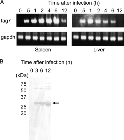
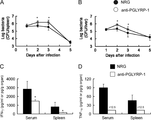
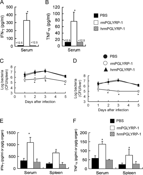
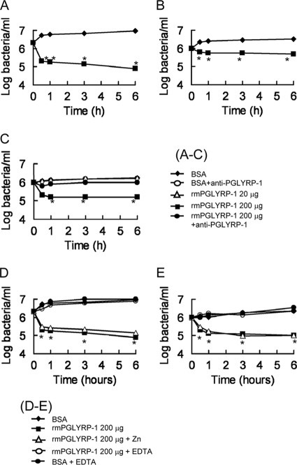
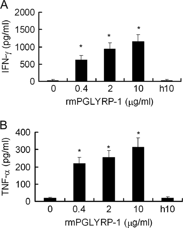
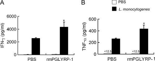
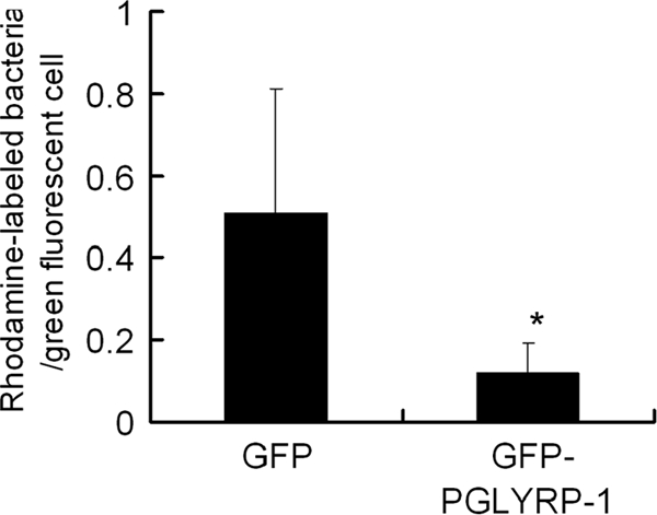
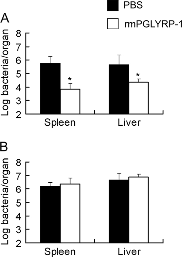
The role of mouse peptidoglycan recognition protein PGLYRP-1 in innate immunity against Listeria monocytogenes infection was studied. The recombinant mouse PGLYRP-1 and a polyclonal antibody specific to PGLYRP-1 were prepared. The mouse PGLYRP-1 showed antibacterial activities against L. monocytogenes and other Gram-positive bacteria. PGLYRP-1 mRNA expression was induced in the spleens and livers of mice infected with L. monocytogenes. The viable bacterial number increased, and the production of cytokines such as gamma interferon (IFN-γ) and tumor necrosis factor alpha (TNF-α) was reduced in mice when mice had been injected with anti-PGLYRP-1 antibody before infection. The levels of IFN-γ and TNF-α titers in the organs were higher and the viable bacterial number was reduced in mice injected with recombinant mouse PGLYRP-1 (rmPGLYRP-1) before infection. PGLYRP-1 could directly induce these cytokines in spleen cell cultures. The elimination of intracellular bacteria was upregulated in NMuLi hepatocyte cells overexpressing PGLYRP-1. The enhancement of the elimination of L. monocytogenes from the organs was observed in IFN-γ(-/-) mice by rmPGLYRP-1 administration but not in TNF-α(-/-) mice. These results suggest that PGLYRP-1 plays a role in innate immunity against L. monocytogenes infection by inducing TNF-α.


Figures

FIG. 1.
FIG. 1.
Expression of PGLYRP-1-coding tag7 gene and PGLYRP-1 protein after L. monocytogenes infection. (A) Mice were infected with 5 × 10⁵ CFU of L. monocytogenes, the spleens and livers were obtained 0, 0.5, 1, 2, 4, 6, and 12 h after infection, and RNA was extracted from the organs. The expression of tag7 and gapdh was estimated by RT-PCR. Representative data are shown for three independent experiments. (B) Mice were infected with 5 × 10⁵ CFU of L. monocytogenes, and the sera were obtained 0, 3, 6, and 12 h after infection. The protein concentration was adjusted to 20 μg/well and applied to Western blotting. Representative data are shown for three independent experiments.
FIG. 2.
FIG. 2.
Inhibition of elimination of L. monocytogenes and endogenous cytokine production by administration of anti-mPGLYRP-1 antibody in mice. C57BL/6 mice were injected with 1 mg of anti-PGLYRP-1 antibody or normal rabbit globulin (NRG) 24 h before infection with 5 × 10⁵ CFU of L. monocytogenes, and viable bacterial numbers in the spleens (A) and livers (B) from mice were determined 1, 2, 3, and 5 days after infection. Similarly, titers of IFN-γ (C) and TNF-α (D) in the sera and spleens 24 h after infection were determined in an anti-PGLYRP-1 antibody-treated group and NRG-treated group. The threshold of detection is indicated by <12.5. Each group consists of six mice. Bars show SD. An asterisk indicates a significant difference from the NRG-treated group at P < 0.05.
FIG. 3.
FIG. 3.
Enhancement of host resistance against L. monocytogenes infection and cytokine production by administration of rmPGLYRP-1. C57BL/6 mice were injected with 100 μg of rmPGLYRP-1, heat-inactivated rmPGLYRP-1, or PBS, and titers of IFN-γ (A) and TNF-α (B) in the sera were determined 24 h after administration. Similarly, C57BL/6 mice were injected with 100 μg of rmPGLYRP-1, heat-inactivated rmPGLYRP-1, or PBS 6 h before infection with 5 × 10⁶ CFU of L. monocytogenes, and viable bacterial numbers in the spleens (C) and livers (D) from mice were determined 1, 2, 3, and 5 days after infection. Titers of IFN-γ (E) and TNF-α (F) in the sera and spleens were determined for the rmPGLYRP-1-treated group, heat-inactivated rmPGLYRP-1-treated group, and PBS-treated group 24 h after infection. The threshold of detection is indicated by <12.5. Each group consists of six mice. Bars show SD. An asterisk indicates a significant difference from the PBS-treated group and heat-inactivated rmPGLYRP-1-treated group at P < 0.05.
FIG. 4.
FIG. 4.
Antibacterial activities of rmPGLYRP-1 against Gram-positive and Gram-negative bacteria. (A to C) An assay mixture for antibacterial activity contained 1 × 10⁶ CFU of S. aureus (A), S. epidermidis (B), and L. monocytogenes (C) rmPGLYRP-1 or bovine serum albumin (BSA) as a control in 1 ml of the assay buffer with or without anti-mPGLYRP-1 antibody (1 mg/ml). After 0.5, 1, 3, and 6 h of incubation, 100 μl of the mixture was taken and plated on tryptic soy agar plates after serial dilution. Each result was obtained from six independent assays. An asterisk indicates that a value is significantly different from the value obtained from BSA controls at P < 0.01. (D, E) An assay mixture for antibacterial activity contained 1 × 10⁶ CFU of S. aureus (D) and L. monocytogenes (E) rmPGLYRP-1 or BSA as a control in 1 ml of the assay buffer with or without 1 mM zinc chloride or 1 mM EDTA. Each result was obtained from six independent assays. An asterisk indicates that a value is significantly different from the value obtained from BSA controls at P < 0.01.
FIG. 5.
FIG. 5.
Cytokine production by stimulation with rmPGLYRP-1 in vitro. (A) Naïve mouse spleen cells were stimulated with 0.4, 2, or 10 μg of rmPGLYRP-1 or 10 μg of heat-inactivated rmPGLYRP-1 for 48 h, and IFN-γ in culture supernatant fluids was assayed. (B) Splenic macrophages were stimulated with 0.4, 2, or 10 μg of rmPGLYRP-1 or 10 μg of heat-inactivated rmPGLYRP-1 for 48 h, and TNF-α in culture supernatant fluids was assayed. h10 means 10 μg of heat-inactivated rmPGLYRP-1. Each group consists of six wells of the plates. Bars show SD. An asterisk indicates that the value is significantly different from that of the unstimulated culture and heat-inactivated rmPGLYRP-1 at P < 0.01.
FIG. 6.
FIG. 6.
Enhancement of cytokine production in spleen cells and macrophages by systemic administration of rmPGLYRP-1. (A) Spleen cells were obtained from C57BL/6 mice 6 h after the injection of 100 μg of rmPGLYRP-1 or PBS in vivo and then were stimulated with PBS or 1 × 10⁷ CFU of viable L. monocytogenes cells for 12 h in vitro. IFN-γ titers in culture supernatant fluids were assayed. (B) Splenic macrophages from C57BL/6 mice pretreated with rmPGLYRP-1 or PBS were stimulated with PBS or viable L. monocytogenes cells for 12 h in vitro. TNF-α titers in culture supernatant fluids were assayed. The threshold of detection is indicated by <12.5. Each group consists of six wells of the plates. Bars show SD. An asterisk indicates that the value is significantly different from that obtained from PBS-pretreated groups at P < 0.01.
FIG. 7.
FIG. 7.
Increased elimination of L. monocytogenes in PGLYRP-1-overexpressing hepatocytes. NMuLi hepatocyte cells seeded at a concentration of 2 × 10⁵ cells/well were transfected with pEGFP-C2/PGLYRP-1 or pEGFP-C2 control vector. Both transfected cells were infected with L. monocytogenes for 30 min, and cell cultivation was continued with the elimination of extracellular bacteria by gentamicin. After 6 h of cultivation, L. monocytogenes cells were labeled with rhodamine, and rhodamine-labeled bacterial numbers in GFP-labeled cells were counted and the ratio of the bacterial number to the cell number was calculated. An asterisk indicates a significant difference from the pEGFP-C2-transfected group at P < 0.05.
FIG. 8.
FIG. 8.
Effect of rmPGLYRP-1 on host resistance against L. monocytogenes infection in IFN-γ−/− mice and TNF-α−/− mice. IFN-γ−/− mice and TNF-α−/− mice were injected with 100 μg of rmPGLYRP-1 or PBS 6 h before infection with 1 × 10⁵ CFU of L. monocytogenes. The number of viable L. monocytogenes cells in organs from IFN-γ−/− mice (A) and TNF-α−/− mice (B) was determined on day 3 after infection. Each group consists of six mice. An asterisk indicates the significant difference from the PBS-treated group at P < 0.01.

References

    1. Bischoff, V., et al. 2004. Function of the drosophila pattern-recognition receptor PGRP-SD in the detection of Gram-positive bacteria. Nat. Immunol. 5:1175-1180. - PubMed
    1. Buchmeier, N. A., and R. D. Schreiber. 1985. Requirement of endogenous interferon-γ production for resolution of Listeria monocytogenes infection. Proc. Natl. Acad. Sci. U. S. A. 82:7404-7408. - PMC - PubMed
    1. Carpenter, S., and L. A. O'Neill. 2007. How important are Toll-like receptors for antimicrobial responses? Cell. Microbiol. 9:1891-1901. - PubMed
    1. Chang, C. I., et al. 2005. Structure of the ectodomain of Drosophila peptidoglycan-recognition protein LCa suggests a molecular mechanism for pattern recognition. Proc. Natl. Acad. Sci. U. S. A. 102:10279-10284. - PMC - PubMed
    1. Cho, S., et al. 2007. Structural insights into the bactericidal mechanism of human peptidoglycan recognition proteins. Proc. Natl. Acad. Sci. U. S. A. 104:8761-8766. - PMC - PubMed

Publication types