. 2011 Feb 25;286(8):6184-91.
doi: 10.1074/jbc.M110.199364. Epub 2010 Dec 6.

Mechanistic insight into human ether-à-go-go-related gene (hERG) K+ channel deactivation gating from the solution structure of the EAG domain

Affiliations

Mechanistic insight into human ether-à-go-go-related gene (hERG) K+ channel deactivation gating from the solution structure of the EAG domain

Frederick W Muskett et al. J Biol Chem. .

Abstract

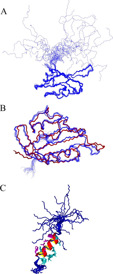
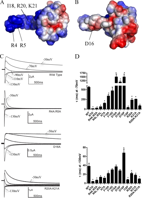
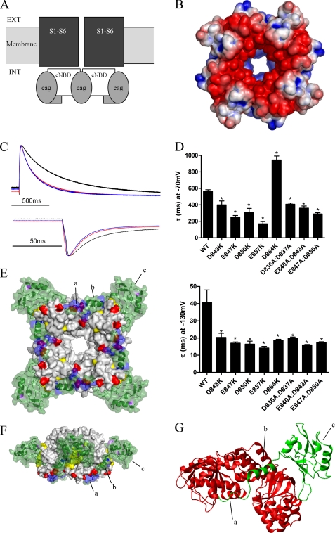
Human ether-à-go-go-related gene (hERG) K(+) channels have a critical role in cardiac repolarization. hERG channels close (deactivate) very slowly, and this is vital for regulating the time course and amplitude of repolarizing current during the cardiac action potential. Accelerated deactivation is one mechanism by which inherited mutations cause long QT syndrome and potentially lethal arrhythmias. hERG deactivation is highly dependent upon an intact EAG domain (the first 135 amino acids of the N terminus). Importantly, deletion of residues 2-26 accelerates deactivation to a similar extent as removing the entire EAG domain. These and other experiments suggest the first 26 residues (NT1-26) contain structural elements required to slow deactivation by stabilizing the open conformation of the pore. Residues 26-135 form a Per-Arnt-Sim domain, but a structure for NT1-26 has not been forthcoming, and little is known about its site of interaction on the channel. In this study, we present an NMR structure for the entire EAG domain, which reveals that NT1-26 is structurally independent from the Per-Arnt-Sim domain and contains a stable amphipathic helix with one face being positively charged. Mutagenesis and electrophysiological studies indicate that neutralizing basic residues and breaking the amphipathic helix dramatically accelerate deactivation. Furthermore, scanning mutagenesis and molecular modeling studies of the cyclic nucleotide binding domain suggest that negatively charged patches on its cytoplasmic surface form an interface with the NT1-26 domain. We propose a model in which NT1-26 obstructs gating motions of the cyclic nucleotide binding domain to allosterically stabilize the open conformation of the pore.


Figures

FIGURE 1.
FIGURE 1.
Structure of the NT1–26 domain of hERG. A, superposition of the protein backbone (Ser26 to Val132) for the family of 20 lowest energy structures calculated using AMBER. B, comparison of the protein backbone of the PAS domain crystal structure (red, Protein Data Bank accession code 1BYW) with the family of NMR structures (blue). C, peptide backbones (blue) of Met1–Gln25 from the 20 NMR structures in A superimposed to illustrate the amphipathic helix extending from Glu11–Gly24. The helix is represented by a ribbon with the hydrophobic residues in magenta and the charged or polar residues in cyan. For clarity, only the side chains of residues in the helix are shown.
FIGURE 2.
FIGURE 2.
A positively charged electrostatic surface on the NT1–26 domain is critical for normal deactivation. A, surface views of the EAG domain of hERG with the residues colored according to their electrostatic potential; areas of significant negative charge are shown in red and significant positive charge in blue and neutral in white. The NT1–26 domain extends out to the left of the molecule and is extensively positively charged on one side. B, 180° rotation of the structure in A about the horizontal axis. Labeled residues in A and B indicate those that when mutated significantly perturb deactivation gating. The position of the Ile18 residue is indicated with an arrow. Arg20 and Lys21 residues are located adjacent and to the right of Ile18 but are obscured in this orientation of the structure. C, representative current traces illustrating differences in rates of deactivation compared with WT hERG for channels in which charged NT1–26 residues have been mutated. Prepulses to +40 mV were applied before stepping down to a range of negative potentials. For clarity, only tail currents at potentials of −50 to −130 mV in 20-mV increments are shown. D, time constants for deactivation at −70 and −130 mV from single exponential fits of tail currents from NT1–26 mutants.* indicates time constants that are significantly different from WT hERG (p < 0.05, n ≥ 5).
FIGURE 3.
FIGURE 3.
Deactivation is regulated by electrostatic interactions between the NT1–26 domain and the cNBD. A, schematic representation of structural domains of the hERG channel, illustrating the intracellular location of the cNBD (and C-linker) with respect to the transmembrane pore and voltage sensor domains (S1–S6). Ext, extracellular; Int, intracellular. B, surface view of the homology model of the cNBD of hERG with residues colored (as in Fig. 2A) according to their electrostatic potential. C, representative current traces (normalized to the peak tail current amplitude) to enable comparison of tail current time courses for WT hERG (black), E847K (red), and E857K (blue). D, time constants for deactivation for cNBD mutant currents, measured as described previously. Most mutations accelerated deactivation at both −70 and −130 mV. D864K was unusual in that deactivation at −70 mV was slowed. Asterisk indicates time constants that are significantly different from WT hERG (p < 0.05, n = 5). E, modeled complex of the EAG domain (green) and the cNBD and C-linker. Four molecules of the EAG domain interact with the tetrameric cNBD. Residues in purple and yellow are those mutated by Al-Owais et al. (23), and residues in red are acidic, and residues in blue are basic. Label a is the unstructured Met1–Pro10 region; b is the amphipathic helix (Gln11–Gly24), and c is the PAS domain. F, modeled complex rotated 90° about the horizontal axis and 45° about the central vertical axis relative to the image in E. G, expanded view of part of modeled complex showing interactions of one EAG domain (green) with two cNBDs (red). The C-linker is not shown. The NT1–26 amphipathic helix (b) sits in a cleft at the interface of two adjacent cNBDs. The unstructured Met1–Pro10 region interacts with the C-helix of one of the cNBDs.

References

    1. Sanguinetti M. C., Jiang C., Curran M. E., Keating M. T. (1995) Cell 81, 299–307 - PubMed
    1. Trudeau M. C., Warmke J. W., Ganetzky B., Robertson G. A. (1995) Science 269, 92–95 - PubMed
    1. Sanguinetti M. C., Jurkiewicz N. K. (1990) J. Gen. Physiol. 96, 195–215 - PMC - PubMed
    1. Sanguinetti M. C., Curran M. E., Spector P. S., Keating M. T. (1996) Proc. Natl. Acad. Sci. U.S.A. 93, 2208–2212 - PMC - PubMed
    1. Curran M. E., Splawski I., Timothy K. W., Vincent G. M., Green E. D., Keating M. T. (1995) Cell 80, 795–803 - PubMed

Publication types

MeSH terms

Substances

Associated data

LinkOut - more resources