. 2011 Feb 25;286(8):6801-7.
doi: 10.1074/jbc.M110.168617. Epub 2010 Dec 6.

The disintegrin-like and cysteine-rich domains of ADAM-9 mediate interactions between melanoma cells and fibroblasts

Affiliations

The disintegrin-like and cysteine-rich domains of ADAM-9 mediate interactions between melanoma cells and fibroblasts

Paola Zigrino et al. J Biol Chem. .

Abstract

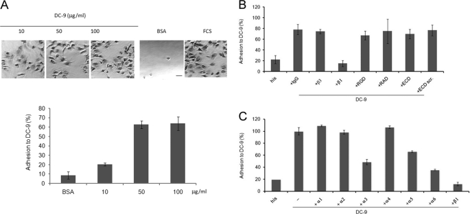
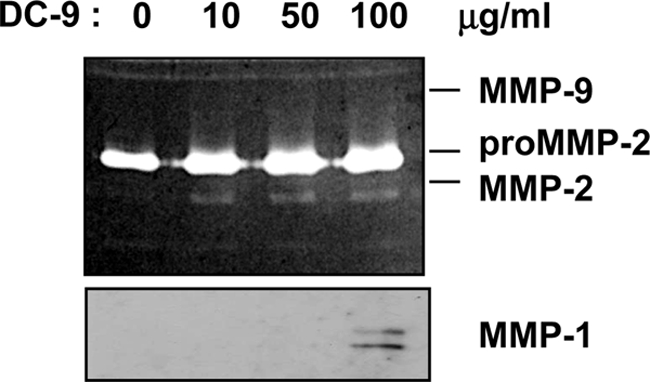
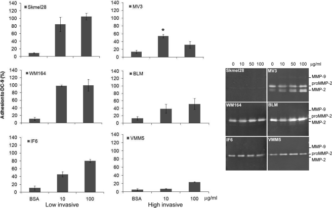
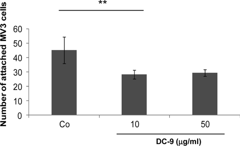
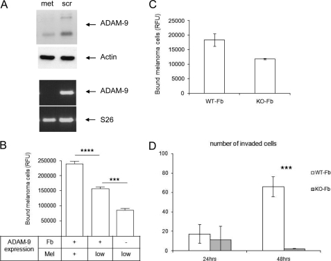
A characteristic of malignant cells is their capacity to invade their surrounding and to metastasize to distant organs. During these processes, proteolytic activities of tumor and stromal cells modify the extracellular matrix to produce a microenvironment suitable for their growth and migration. In recent years the family of ADAM proteases has been ascribed important roles in these processes. ADAM-9 is expressed in human melanoma at the tumor-stroma border where direct or indirect interactions between tumor cells and fibroblasts occur. To analyze the role of ADAM-9 for the interaction between melanoma cells and stromal fibroblasts, we produced the recombinant disintegrin-like and cysteine-rich domain of ADAM-9 (DC-9). Melanoma cells and human fibroblasts adhered to immobilized DC-9 in a Mn(2+)-dependent fashion suggesting an integrin-mediated process. Inhibition studies showed that adhesion of fibroblasts was mediated by several β1 integrin receptors independent of the RGD and ECD recognition motif. Furthermore, interaction of fibroblasts and high invasive melanoma cells with soluble recombinant DC-9 resulted in enhanced expression of MMP-1 and MMP-2. Silencing of ADAM-9 in melanoma cells significantly reduced cell adhesion to fibroblasts. Ablation of ADAM-9 in fibroblasts almost completely abolished these cellular interactions and melanoma cell invasion in vitro. In summary, these results suggest that ADAM-9 expression plays an important role in mediating cell-cell contacts between fibroblasts and melanoma cells and that these interactions contribute to proteolytic activities required during invasion of melanoma cells.


Figures

FIGURE 1.
FIGURE 1.
Fibroblasts adhere to the adhesive domains of ADAM-9. A, microtiter plates were coated with increasing concentrations of recombinant His-tagged DC-9 protein as indicated. Coating with BSA (1% heat inactivated) or FCS were used as negative and positive control, respectively. Microphotographs were taken after fixation of the cells (scale bar 10 μm). B and C, microtiter plates were coated with His-tagged DC-9 protein (10 μg/ml) or his peptide (0.6 μm). Fibroblasts were incubated in assay buffer in the presence of 10 μg/ml inhibitory antibodies as indicated. IgG isotype control or 0.6 μm of each RGD, RAD (control), ECD or scrambled-ECD (scr.; control) peptide was added before plating. Adhesion is expressed as percentage of adhesion determined on FCS, which was arbitrarily set as 100%. In B, representative results of four independent experiments, each performed in triplicates; in C representative results of three independent experiments, each performed in triplicates.
FIGURE 2.
FIGURE 2.
Adhesion of fibroblasts to DC-9 alters MMP-1 and -2 expression. Primary human fibroblasts were starved for 24 h in serum-free medium before incubation with DC-9 protein (10, 50, 100 μg/ml). After stimulation, supernatants were collected and analyzed by gelatin zymography. The 72 kDa inactive form and the 62/59 kDa active forms of MMP-2 are shown in the zymogram (upper panel). MMP-1 in supernatants was detected by Western blotting using rabbit-anti-MMP-1 antibodies. The antibodies detect two bands of 57 and 52 kDa.
FIGURE 3.
FIGURE 3.
Melanoma cells of different invasive potential adhere to DC-9. Melanoma cells were seeded on microtiter plates coated with 10 and 100 μg/ml of DC-9 protein for 1 h. Coating with BSA (1% heat-inactivated) and FCS were used as negative and positive controls, respectively. Adhesion is expressed as percentage of adhesion to FCS, which was set arbitrarily as 100%. For gelatin zymography melanoma cells were starved for 24 h in serum-free medium before stimulation with DC-9 protein (10, 50, 100 μg/ml). Then the supernatants were collected and analyzed by gelatin zymography. The 72 kDa inactive form and the 62/59 kDa active forms of MMP-2 are indicated in the zymogram. Significance of MV3 adhesion as compared with SKmel23 and WM164, *, p < 0,05.
FIGURE 4.
FIGURE 4.
The DC domain of ADAM-9 antagonise fibroblast-melanoma cell interaction. MV3 melanoma cells, labeled with the fluorochrome CMFDA, were preincubated in suspension with either 0.6 μm his peptide (Co) or 10 and 50 μg/ml of recombinant, His-tagged DC-9 protein before seeding on human primary fibroblasts. After 1 h, attached cells were counted in 5 microscopic fields of 3 separate wells. The bars represent the mean ± S.D. of cells attached per field. **, p < 0,006.
FIGURE 5.
FIGURE 5.
Silencing of ADAM-9 in MV3 melanoma cells results in reduced melanoma-fibroblast interaction. A, MV3 cells were transiently transfected with ADAM-9 siRNA (met) or control siRNA (scr). After 48 h, ADAM-9 mRNA levels were determined by RT-PCR. S26 levels were used for normalization. Protein expression was analyzed in cell lysates by Western blotting, and detection of actin was used as loading control. B, 48 h after transfection of MV3 cells (Mel) with ADAM-9 (low) or control siRNA (+), the MV3 cells were labeled with CMFDA and seeded on confluent fibroblast monolayers (Fb) derived from either wild type (+) or ADAM-9-deficient (−) mice. After 20 min, bound cells were quantified by fluorimetric analysis. Bars represent mean ± S.D. of the relative fluorescence units of three independent experiments. ****, p < 0.0001; ***, p < 0.0004. C, B16F1 murine melanoma cells were labeled with CMFDA and seeded on confluent fibroblast monolayers derived from either wild type (WT-Fb) or ADAM-9-deficient (KO-Fb) mice. Numbers of bound cells were quantified after 20 min. *, p < 0,006. D, invasion of GFP-transfected B16F1 melanoma cells was determined in transwell cultures with wild type (WT) or ADAM-9-deficient (KO) fibroblasts. Melanoma cells (mel; 5 × 10⁵/ml) suspended in a 2:1 ratio with fibroblasts in 0.5-ml serum-free RPMI medium were filled in the upper compartment, while fibroblast-conditioned medium was used as a chemoattractant in the lower compartment. After 24 and 48 h, the numbers of GFP-positive melanoma cells that had migrated through the matrigel to the lower side of the filter were counted. Bars represent the mean ± S.D. of the cell numbers determined per filter in three independent experiments. ***, p < 0,0001.

References

    1. Edwards D. R., Handsley M. M., Pennington C. J. (2008) Mol. Aspects Med. 29, 258–289 - PMC - PubMed
    1. Bergers G., Coussens L. M. (2000) Curr. Opin. Genet. Dev. 10, 120–127 - PubMed
    1. Schlöndorff J., Blobel C. P. (1999) J. Cell Sci. 112, 3603–3617 - PubMed
    1. Zhang X. P., Kamata T., Yokoyama K., Puzon-McLaughlin W., Takada Y. (1998) J. Biol. Chem. 273, 7345–7350 - PubMed
    1. Chen M. S., Tung K. S., Coonrod S. A., Takahashi Y., Bigler D., Chang A., Yamashita Y., Kincade P. W., Herr J. C., White J. M. (1999) Proc. Natl. Acad. Sci. U.S.A. 96, 11830–11835 - PMC - PubMed

Publication types

MeSH terms