. 2010 Dec 13;191(6):1079-88.
doi: 10.1083/jcb.201006083. Epub 2010 Dec 6.

Vesicle trafficking maintains nuclear shape in Saccharomyces cerevisiae during membrane proliferation

Affiliations

Vesicle trafficking maintains nuclear shape in Saccharomyces cerevisiae during membrane proliferation

Micah T Webster et al. J Cell Biol. .

Abstract

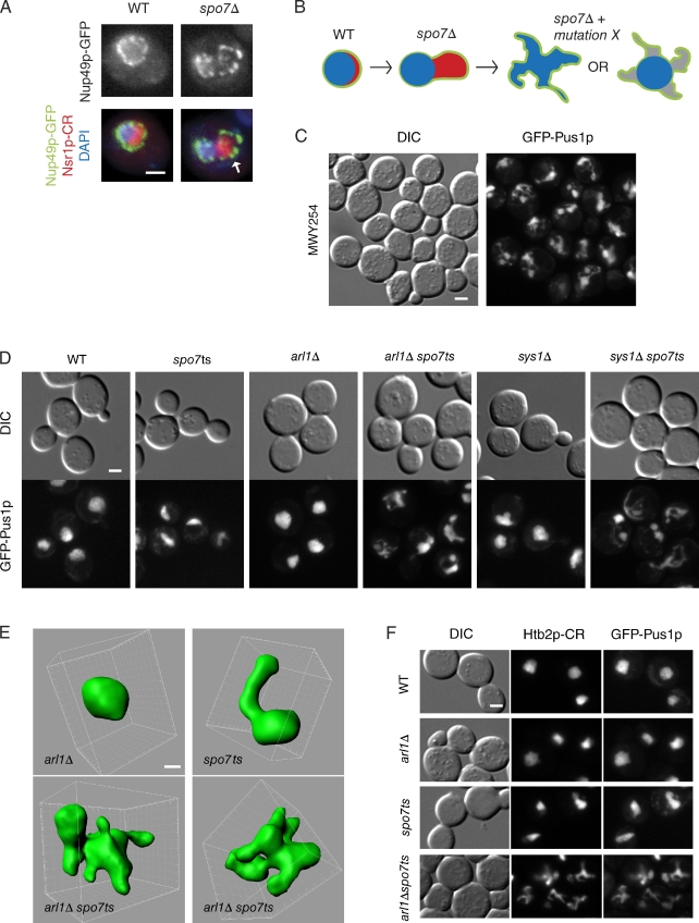
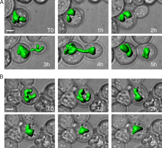
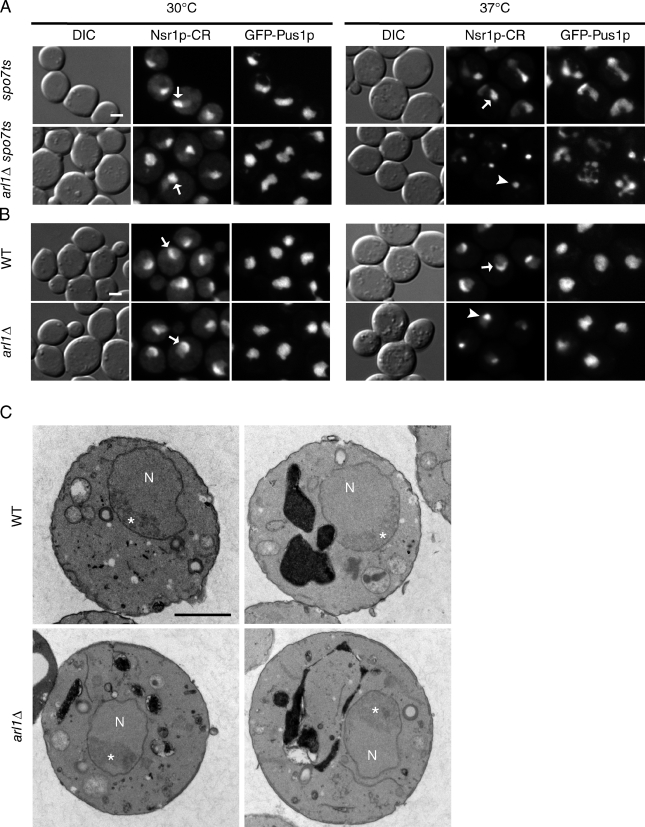
The parameters that control nuclear size and shape are poorly understood. In yeast, unregulated membrane proliferation, caused by deletion of the phospholipid biosynthesis inhibitor SPO7, leads to a single nuclear envelope "flare" that protrudes into the cytoplasm. This flare is always associated with the asymmetrically localized nucleolus, which suggests that the site of membrane expansion is spatially confined by an unknown mechanism. Here we show that in spo7Δ cells, mutations in vesicle-trafficking genes lead to multiple flares around the entire nucleus. These mutations also alter the distribution of small nucleolar RNA-associated nucleolar proteins independently of their effect on nuclear shape. Both single- and multi-flared nuclei have increased nuclear envelope surface area, yet they maintain the same nuclear/cell volume ratio as wild-type cells. These data suggest that, upon membrane expansion, the spatial confinement of the single nuclear flare is dependent on vesicle trafficking. Moreover, flares may facilitate maintenance of a constant nuclear/cell volume ratio in the face of altered membrane proliferation.


Figures

Figure 1.
Figure 1.
ARL1 and SYS1 affect nuclear morphology in spo7Δ cells. (A) The flare phenotype in spo7Δ cells. Images of fixed wild-type cells (WT) and spo7Δ cells are shown. The flare in spo7Δ cells is marked with an arrow. The NE is identified by the nucleoporin Nup49p fused to GFP (Nup49p-GFP); the nucleolus is identified by the nucleolar protein Nsr1p fused to mCherry red fluorescent protein (Nsr1p-CR); the DNA is stained with DAPI. (B) Diagram of the yeast nucleus showing the NE (green), the nucleolus (red), and the DNA (blue) in wild-type and spo7Δ cells, and hypothetical nuclear phenotypes that would result from a mutation leading to a multi-flare phenotype with (right) or without (left) loss of DNA tethering to the NE. (C) Nuclear phenotype of strain MWY254, carrying a mutation that is synthetically lethal with spo7Δ, observed after a 2-h temperature shift to 37°C. Nuclear morphology was assessed with the nucleoplasmic protein Pus1p fused to GFP (GFP-Pus1p). For comparison to nuclei of wild-type and spo7ts cells, see D or F. (D) Nuclear phenotypes by GFP-Pus1p, associated with arl1Δ and sys1Δ mutations, alone or in combination with spo7ts, after a 2-h temperature shift to 37°C. (E) Three-dimensional reconstruction of nuclei from arl1Δ (top left), spo7ts (top right), and two arl1 spo7ts (bottom) cells. Cells were shifted to 37°C for 2 h. (F) Spatial distribution of chromatin in wild-type, arl1Δ, spo7ts, and arl1Δ spo7ts strains. Chromatin is visualized by the histone H2B fused to mCherry (Htb2p-CR), and nuclear morphology is detected by GFP-Pus1p. Cells were shifted to 37°C for 2 h. Bars: (A, C, D, and F) 2 µm; (E) 1 µm.
Figure 2.
Figure 2.
arl1Δ spo7ts cells with multi-flared nuclei can recover and divide. 1-h time points of live arl1Δ spo7ts cells grown at 30°C after a 3-h temperature shift to 37°C. Three-dimensional reconstructions of nuclei (with GFP-Pus1) are overlaid on corresponding phase images of cells. (A) A cell with a multi-flared nucleus at t0 divides over the 5-h time course. Mother and daughter cells are seen at the 5-h time point, each with its individual nucleus. (B) A cell with a multi-flared nucleus at t0 does not divide, but undergoes dynamic shape changes. Bars, 2 µm.
Figure 3.
Figure 3.
Deleting ARL1 causes the coalescence of Nsr1p-CR to a single focus but does not affect overall nucleolar structure. (A) spo7ts cells, with and without ARL1, were fixed either before (left) or after a 3-h temperature shift to 37°C (right), then examined for nuclear morphology (with GFP-Pus1p) and Nsr1p-CR distribution. Arrows show examples of Nsr1p-CR in single flares; the arrowhead shows an example of focal Nsr1p-CR. (B) Wild-type (WT) and arl1Δ strains were treated the same as in A. Arrows show examples of Nsr1p-CR in crescent form; the arrowhead shows an example of focal Nsr1p-CR. (C) Electron micrographs of wild-type and arl1Δ cells fixed after a 2-h temperature shift to 37°C. Nucleoli (labeled with asterisks) are the darker, more electron-dense regions within the nuclei (N). Bars, 2 µm.

References

    1. Abudugupur A., Mitsui K., Yokota S., Tsurugi K. 2002. An ARL1 mutation affected autophagic cell death in yeast, causing a defect in central vacuole formation. Cell Death Differ. 9:158–168 10.1038/sj.cdd.4400942 - DOI - PubMed
    1. Behnia R., Panic B., Whyte J.R., Munro S. 2004. Targeting of the Arf-like GTPase Arl3p to the Golgi requires N-terminal acetylation and the membrane protein Sys1p. Nat. Cell Biol. 6:405–413 10.1038/ncb1120 - DOI - PubMed
    1. Belgareh N., Doye V. 1997. Dynamics of nuclear pore distribution in nucleoporin mutant yeast cells. J. Cell Biol. 136:747–759 10.1083/jcb.136.4.747 - DOI - PMC - PubMed
    1. Bender A., Pringle J.R. 1991. Use of a screen for synthetic lethal and multicopy suppressee mutants to identify two new genes involved in morphogenesis in Saccharomyces cerevisiae. Mol. Cell. Biol. 11:1295–1305 - PMC - PubMed
    1. Boeke J.D., LaCroute F., Fink G.R. 1984. A positive selection for mutants lacking orotidine-5′-phosphate decarboxylase activity in yeast: 5-fluoro-orotic acid resistance. Mol. Gen. Genet. 197:345–346 10.1007/BF00330984 - DOI - PubMed

Publication types

MeSH terms

Substances