. 2010 Dec 13;191(6):1089-95.
doi: 10.1083/jcb.201007030. Epub 2010 Dec 6.

CDK5RAP2 stimulates microtubule nucleation by the gamma-tubulin ring complex

Affiliations

CDK5RAP2 stimulates microtubule nucleation by the gamma-tubulin ring complex

Yuk-Kwan Choi et al. J Cell Biol. .

Abstract

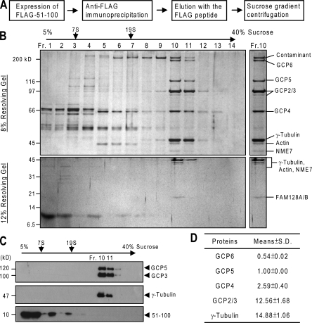
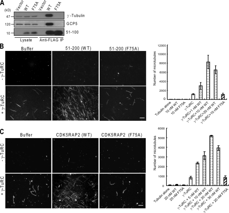
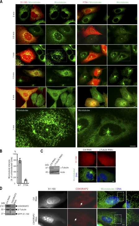
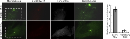
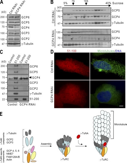
CDK5RAP2 is a human microcephaly protein that contains a γ-tubulin complex (γ-TuC)-binding domain conserved in Drosophila melanogaster centrosomin and Schizosaccharomyces pombe Mto1p and Pcp1p, which are γ-TuC-tethering proteins. In this study, we show that this domain within CDK5RAP2 associates with the γ-tubulin ring complex (γ-TuRC) to stimulate its microtubule-nucleating activity and is therefore referred to as the γ-TuRC-mediated nucleation activator (γ-TuNA). γ-TuNA but not its γ-TuC-binding-deficient mutant stimulates microtubule nucleation by purified γ-TuRC in vitro and induces extensive, γ-TuRC-dependent nucleation of microtubules in a microtubule regrowth assay. γ-TuRC bound to γ-TuNA contains NME7, FAM128A/B, and actin in addition to γ-tubulin and GCP2-6. RNA interference-mediated depletion of CDK5RAP2 impairs both centrosomal and acentrosomal microtubule nucleation, although γ-TuRC assembly is unaffected. Collectively, these results suggest that the γ-TuNA found in CDK5RAP2 has regulatory functions in γ-TuRC-mediated microtubule nucleation.


Figures

Figure 1.
Figure 1.
Isolation of γ-TuCs bound to γ-TuNA. (A) Schematic outline of the isolation procedure. (B) After gradient centrifugation, an aliquot of each fraction was resolved by SDS-PAGE followed by silver staining. Proteins resolved from the peak fraction of γ-TuRC (Fr. 10) were identified by mass spectrometry. The contaminant protein above GCP6 also appeared in the precipitates of blank beads. (C) The gradient fractions were analyzed by immunoblotting. (D) In a replicate gel stained with Sypro ruby, the relative amounts of γ-tubulin and GCPs were determined from the isolated γ-TuRC (Fr. 10) to derive the stoichiometry. The ratios of proteins to GCP5 are presented as mean ± SD from three independent experiments.
Figure 2.
Figure 2.
Stimulation of γ-TuRC for microtubule nucleation. (A) After immunoprecipitation of ectopically expressed proteins through the tag moiety (i.e., Flag), the immunoprecipitates and cell lysates were examined for γ-tubulin and GCP5. WT, 51–100 wild type; F75A, 51–100 (F75A). (B and C) Microtubule polymerization was performed with or without the isolated γ-TuRC and 51–200 (B) or the entire CDK5RAP2 protein (C). Representative microscopic fields of polymerized microtubules are shown. Microtubules were counted from 20 random fields to derive the mean numbers of microtubules. Data are shown as mean ± SD of three independent experiments. Bar, 10 µm.
Figure 3.
Figure 3.
Microtubule nucleation induced by γ-TuNA in transfected cells. (A and B) Microtubule regrowth was performed on U2OS cells expressing 51–100 or its F75A mutant. (bottom) Enlarged views of the boxed areas are shown. The microtubule intensities of regrowth for 1 min were quantified from cells expressing the proteins at similar levels and were expressed relative to untransfected cells (B). At least 20 cells were measured in each of three independent experiments. (B) Error bars indicate mean ± SD. (C) Microtubule regrowth (1 min) was performed after γ-tubulin was depleted by RNAi. (left) The expressions of γ-tubulin and actin were examined by immunoblotting. (right) Cells transfected with 51–100 were subjected to the regrowth assay. The depletion of γ-tubulin reduced microtubule regrowth to 9.20 ± 2.89% of control cells (n = 60 cells for each quantification). (D) 51–100 (GFP tagged) was expressed in the background of endogenous CDK5RAP2 depleted by RNAi or in the control. (left) Cell extracts were immunoblotted for GFP-51–100 (anti-GFP), endogenous CDK5RAP2, and β-tubulin. (right) The cells were stained for microtubules (anti–α-tubulin) and endogenous CDK5RAP2 after microtubule regrowth for 1 min. Arrows denote centrosomes. (right) Enlarged views of the boxed areas are shown. Bars, 10 µm.
Figure 4.
Figure 4.
Cytoplasmic microtubule nucleation is impaired by the depletion of CDK5RAP2. (left) Microtubules regrew for 1.5 min after depolymerization in MRC-5 cells. Asterisks mark acentrosomal microtubules. Enlarged views of the boxed areas are shown. Bar, 10 µm. (right) The bar graph shows the number of acentrosomal microtubules. Data are shown as mean ± SD of three independent experiments; at least 20 cells were analyzed in each experiment.
Figure 5.
Figure 5.
Depletion of GCP4 disrupts the association of γ-TuNA with γ-TuRC. (A) siRNA-transfected HEK293T extracts were immunoblotted for γ-tubulin and GCPs. (B) Cell extracts were subjected to sucrose gradient centrifugation. An aliquot of each gradient fraction was analyzed by immunoblotting. (C) His-Flag–CDK5RAP2 (51–200) was used in the pull down of γ-TuCs from siRNA-transfected extracts. The anti-Flag precipitates were analyzed by immunoblotting. Note that the protein band below GCP4 is a heat shock protein bound nonspecifically to anti-Flag beads. (D) Microtubule regrowth was performed on U2OS cells with the regrowth time of 1 min. GCP4 depletion reduced microtubule regrowth to 14.02 ± 3.84% of control cells (n = 60 cells for each quantification). Bar, 10 µm. (E) Model for the activation of γ-TuRC–mediated microtubule nucleation by γ-TuNA. After γ-TuRC assembly, γ-TuNA binds to the complex to stimulate its nucleation of microtubules.

References

    1. Bond J., Roberts E., Springell K., Lizarraga S.B., Lizarraga S., Scott S., Higgins J., Hampshire D.J., Morrison E.E., Leal G.F., et al. 2005. A centrosomal mechanism involving CDK5RAP2 and CENPJ controls brain size. Nat. Genet. 37:353–355 10.1038/ng1539 - DOI - PubMed
    1. Fava F., Raynaud-Messina B., Leung-Tack J., Mazzolini L., Li M., Guillemot J.C., Cachot D., Tollon Y., Ferrara P., Wright M. 1999. Human 76p: a new member of the γ-tubulin–associated protein family. J. Cell Biol. 147:857–868 10.1083/jcb.147.4.857 - DOI - PMC - PubMed
    1. Fong K.W., Choi Y.K., Rattner J.B., Qi R.Z. 2008. CDK5RAP2 is a pericentriolar protein that functions in centrosomal attachment of the γ-tubulin ring complex. Mol. Biol. Cell. 19:115–125 10.1091/mbc.E07-04-0371 - DOI - PMC - PubMed
    1. Fong K.W., Hau S.Y., Kho Y.S., Jia Y., He L., Qi R.Z. 2009. Interaction of CDK5RAP2 with EB1 to track growing microtubule tips and to regulate microtubule dynamics. Mol. Biol. Cell. 20:3660–3670 10.1091/mbc.E09-01-0009 - DOI - PMC - PubMed
    1. Haren L., Remy M.H., Bazin I., Callebaut I., Wright M., Merdes A. 2006. NEDD1-dependent recruitment of the γ-tubulin ring complex to the centrosome is necessary for centriole duplication and spindle assembly. J. Cell Biol. 172:505–515 10.1083/jcb.200510028 - DOI - PMC - PubMed

Publication types

Substances