. 2011 Jan 1;186(1):602-13.
doi: 10.4049/jimmunol.1002850. Epub 2010 Dec 6.

Critical modifier role of membrane-cystic fibrosis transmembrane conductance regulator-dependent ceramide signaling in lung injury and emphysema

Affiliations

Critical modifier role of membrane-cystic fibrosis transmembrane conductance regulator-dependent ceramide signaling in lung injury and emphysema

Manish Bodas et al. J Immunol. .

Abstract

Ceramide accumulation mediates the pathogenesis of chronic obstructive lung diseases. Although an association between lack of cystic fibrosis transmembrane conductance regulator (CFTR) and ceramide accumulation has been described, it is unclear how membrane-CFTR may modulate ceramide signaling in lung injury and emphysema. Cftr(+/+) and Cftr(-/-) mice and cells were used to evaluate the CFTR-dependent ceramide signaling in lung injury. Lung tissue from control and chronic obstructive pulmonary disease patients was used to verify the role of CFTR-dependent ceramide signaling in pathogenesis of chronic emphysema. Our data reveal that CFTR expression inversely correlates with severity of emphysema and ceramide accumulation in chronic obstructive pulmonary disease subjects compared with control subjects. We found that chemical inhibition of de novo ceramide synthesis controls Pseudomonas aeruginosa-LPS-induced lung injury in Cftr(+/+) mice, whereas its efficacy was significantly lower in Cftr(-/-) mice, indicating that membrane-CFTR is required for controlling lipid-raft ceramide levels. Inhibition of membrane-ceramide release showed enhanced protective effect in controlling P. aeruginosa-LPS-induced lung injury in Cftr(-/-) mice compared with that in Cftr(+/+) mice, confirming our observation that CFTR regulates lipid-raft ceramide levels and signaling. Our results indicate that inhibition of de novo ceramide synthesis may be effective in disease states with low CFTR expression like emphysema and chronic lung injury but not in complete absence of lipid-raft CFTR as in ΔF508-cystic fibrosis. In contrast, inhibiting membrane-ceramide release has the potential of a more effective drug candidate for ΔF508-cystic fibrosis but may not be effectual in treating lung injury and emphysema. Our data demonstrate the critical role of membrane-localized CFTR in regulating ceramide accumulation and inflammatory signaling in lung injury and emphysema.


Figures

Figure 1
Figure 1. CFTR regulates innate and adaptive immune responses
The macrophages and neutrophils derived from Cftr−/− mice show significant increase in constitutive (A) IL-6 and (B) MPO (myeloperoxidase levels, only in neutrophils) secretion in the culture supernatants as compared to the Cftr+/+, ***p<0.001. (C) The bronchoalveolar lavage fluid (BALF) from Cftr−/− mice show significant increase in (*p<0.05) in the basal and Pa-LPS (20μg i.t., 24 hours) induced MPO levels as compared to the Cftr+/+. (D) The splenocytes from Cftr−/− mice show significantly (***p<0.001, **p<0.01) higher Concanavalin A (ConA, 5 or 10 μg/ml) induced cell proliferation as compared to the Cftr+/+ mice. (E) The culture supernatants from these splenocytes (D) have significantly (***p<0.001) higher IL-6 levels in the Cftr−/− as compared to the Cftr+/+. (F) The flow cytometry analysis shows Cftr expression in CD4+ Cftr+/+ mice splenocytes (i) while Cftr−/− splenocytes were used as a negative control. The significant increase in percentage of CD4+IFNγ+ (ii) and CD4+FoxP3+ (iii) cells in the Cftr−/− splenocytes as compared to the Cftr+/+ is indicative of the constitutive T cell activation in the absence of CFTR. (G) Immunofluorescence staining verifies the increase in constitutive and Pa-LPS induced FoxP3 expression and nuclear localization in Cftr−/− mice lungs as compared to the Cftr+/+ (Scale: 50 μm). (H) Differences in basal and Con A (5μg/ml) induced FoxP3 expression in Cftr+/+ and Cftr−/− splenocytes is confirmed by western blotting. β-actin blot shows the equal loading. (I) Densitometry analysis of FoxP3 expression (in H) normalized to β- actin. Data represent n=3 in each group and error bars depict mean ± SEM.
Figure 2
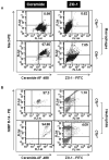
Figure 2. Ceramide and zona occludens 1 (ZO-1) expression is elevated in Cftr−/− mice immune cells
Flow cytometry analysis showing ZO-1 and ceramide expression in macrophages (A) and neutrophils (B) from Cftr+/+ and Cftr−/− mice. Thioglycollate-elicited peritoneal macrophages and neutrophils were immunostained for Mac-3 (macrophage) and NIMP-R14 (neutrophil) markers and co-staining with ceramide (left panel) or ZO-1 (right panel) antibodies was used to quantify the percentage changes in the number of positive cells. The upper right quadrant shows the percentage gated cells positive for both the primary antibodies as indicated. Data from n=3 mice shows an very significant increase in ceramide positive cells (left panel) in Cftr−/− mice (97.85%) derived macrophages (A) as compared to the Cftr+/+ (0.99%) while neutrophils (B) have no change. In contrast, expression of lipid-raft marker, ZO-1 (right panel) shows a significant increase in both the cell types (A&B) in the absence of CFTR (Cftr−/−) indicating the role of CFTR in tight-junction formation.
Figure 3
Figure 3. Severity of inflammatory lung disease inversely correlates with the membrane-CFTR levels
(A) Human lung tissue sections from each group at Gold- 0 (at risk), I (mild), II (moderate) and III-IV (severe and very-severe) COPD (n=4-10) were stained with H&E (bottom panel) showing a significant increase in inflammatory cells and emphysema in moderate and severe COPD as compared to the mild. The lung tissue sections immunostained with CFTR (green, upper panel) or ceramide (green, 3rd panel) show significant decrease in membrane CFTR expression at advanced stage of COPD lung disease while ceramide levels increase. Nuclear (Hoechst) staining is shown in blue (Scale: white-50 μm, red-10 μm, black-100 μm). (B) Densitometric analysis confirms the statistical significance (p< 0.001) and illustrates the correlation of CFTR and ceramide expression with severity of lung emphysema. (C) The HEK-293 cells transfected with wt-CFTR and treated with increasing doses of cigarette smoke extract (CSE) for 12 hours (n=3) show an inverse relationship between increasing CSE dose and expression of membrane CFTR (mature C-band, left panel). The total cell lysates from HEK-293 cells either control (a) or transfected with wt-CFTR (b) shows the absence of CFTR (B and C bands) in the control cells (right panel). (D) The lung lysates from air and CS exposed mice (n=3) were used for either Cftr immunoprecipitation (CFTR-169, upper panel) or lipid-raft isolation and Cftr protein levels were detected by western blotting. The data shows a significant decrease in membrane and lipid-raft Cftr protein expression in the lungs of CS exposed mice. (E) Densitometry analysis of membrane- and raft- Cftr expression from control and CS groups (in D) is shown as mean ± SEM of triplicate samples (**p<0.01, ***p<0.001). (F) The longitudinal lung sections from air or CS exposed mice (same experiment as D) show an increased ceramide and ZO-1 co-staining (red arrow) in the CS exposed lungs verifying that CS modulates lipid-raft and ceramide signaling in murine lungs.
Figure 4
Figure 4. CFTR regulates de novo and membrane ceramide signaling
Bronchoalveolar lavage fluids (BALF) from 3-5 C57BL/6 Cftr+/+ or Cftr−/− mice, treated intratracheally with PBS (control), Pa-LPS (20 μg/mouse; 12 hrs), Fumonisin B1 (FB1, 50 μg/mouse; 24 hrs) and/or Amitriptyline (AMT, 50 μg/mouse; 24 hrs) were used to quantify the IL-6 and IL-1β levels. (A) Inhibition of de novo ceramide synthesis by FB1 treatment significantly decreases Pa-LPS induced IL-6 and IL-1β (*p<0.05, **p<0.001) in Cftr+/+ mice (i, ii), but FB1 has a modest effect on Pa-LPS induced IL-6 levels in Cftr−/− mice (iii, iv). (B) Inhibition of membrane ceramide release by AMT treatment is relatively less protective against Pa-LPS induced lung injury in Cftr+/+ mice (i, ii), but effectively controls the inflammatory cytokines in Cftr−/− mice (iii, iv). The data shows that inhibition of de novo and membrane ceramide release can control Pa-LPS induced lung injury in the presence or absence of CFTR, respectively. This also indicates that CFTR can regulate de novo and membrane ceramide signaling. Data represent the averages of triplicate ELISAs from n=3-5 samples and is shown as mean ± SEM; *p<0.05, **p<0.01, ***p<0.001).
Figure 5
Figure 5. CFTR regulates lipid-raft expression and signaling via ceramide
(A) CFBE4lo-wt-CFTR (wt-CFBE) and CFBE41o- cells were stimulated with Pa-LPS (10 ng/ml) or Fumonisin B1 (FB1, 50μM) for 24 hours. The lipid-raft protein extracts were isolated from these cells and expression of lipid-raft marker, ZO-2 was quantified by western blotting. Data show significant downregulation (>2 fold) of lipid-raft ZO-2 expression with Pa-LPS or FB1 treatment only in the presence of wt-CFTR indicating that Cftr is a critical regulator of Pa-LPS or ceramide mediated lipid-raft expression and signaling. The same membrane was blotted with α-actin as a loading control. (B) Immunostaining for ZO-2 shows its increased expression in lung tissue sections from Cftr−/− mice (n=3) as compared to the Cftr+/+ (n=3) (upper panel) verifying that CFTR regulates the expression of lipid-raft protein, ZO-2. (C) The lung sections from Cftr+/+ and Cftr−/− mice (n=4-5), treated with PBS or Pa-LPS (20 μg/mouse; 24 hrs), immunostained for ZO-1 (green) and ceramide (red) show significant increase in constitutive and Pa-LPS induced ZO-1 and ceramide levels (upper panel) in Cftr−/− as compared to the Cftr+/+s. The co-localization of ceramide with ZO-1 verifies the lipid-raft localization of ceramide in the absence of CFTR. The CFTR immunostaining (green, middle panel) shows the CFTR expression levels in the Cftr+/+ mice lungs while Cftr−/− are shown as a negative control. (D) The densitometry and spearman’s correlation coefficient analysis of ZO-1 and ceramide staining (C) shows the statistical significance of immunostaining data. (Scale: white bar- 50 μm, red bar- 10 μm, black-100 μm).
Figure 6
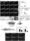
Figure 6. The PDZ-interacting domain of CFTR regulates ceramide accumulation. (A)
The HEK-293 cells were transiently transfected with pEGFP WT- or ΔTRL- CFTR plasmid constructs and one experimental group was treated with 100 μg/ml CSE for 12 hours. The cells were stained and analyzed for ceramide (R-PE, FL-2) and GFP expression (FL-1) by flow cytometry. The data represents three independent experiments. Expression of CFTR lacking the PDZ-interacting domain shows an increase in basal (49.85% to 56.48%) and CSE induced ceramide accumulation (69.22% to 80.56%), indicating towards the crucial role of PDZ-binding domains in regulating CFTR-dependent ceramide signaling. (B) The HEK-293 cells transiently over-expressing WT- or ΔTRL- CFTR plasmids (n=3) were incubated with FITC-labeled E. coli LPS for 3 hours and analyzed by flow cytometry (unpermeabilized cells). The transient expression of CFTR lacking the PDZ-binding domain results in reduced binding of LPS to the plasma membrane. We anticipate that less LPS binding to ΔTRL expressing cells is a direct consequence of its reduced cell surface expression and/or lipid-raft translocation. (C) The lipid-raft proteins from HEK 293 cells expressing WT- or ΔTRL- CFTR were analyzed for CFTR expression by western blotting (a, 30sec and b, 20 min exposure). The data shows that lack of the PDZ-interacting domain of CFTR compromises its membrane expression (b, left panel) and translocation to the lipid-rafts (a&b, right panel). (D) Densitometry analysis of membrane- and raft- CFTR expression from WT and ΔTRL- CFTR groups (in C).
Figure 7
Figure 7. Schematic of CFTR mediated ceramide signaling
Schematic illustrates the critical role of lipid-raft CFTR in controlling ceramide (sphingomyelin) and inflammatory (TNFα) or apoptotic (CD95) signaling. Our model predicts that the absence or decrease in lipid-raft CFTR expression culminates these regulatory functions, resulting in NFκB mediated hyper-inflammatory response. Environmental factors such as Pseudomonas aeruginosa infection or cigarette smoke exposure further exaggerate the lipid-raft signaling and contribute to the pathogenesis of chronic inflammatory or apoptotic signaling by modulating CFTR lipid-raft expression that controls ceramide accumulation. We anticipate that in the absence of lipid-raft CFTR, membrane ceramide accumulation induces lipid-raft fusion and large scale clustering of the membrane receptors that result in lung injury and emphysema.

References

    1. Wedzicha JA, Donaldson GC. Exacerbations of chronic obstructive pulmonary disease. Respir Care. 2003;48:1204–1213. discussion 1213-1205. - PubMed
    1. Heijerman H. Infection and inflammation in cystic fibrosis: a short review. J Cyst Fibros. 2005;4(Suppl 2):3–5. - PubMed
    1. Martin RJ. Infections and asthma. Clin Chest Med. 2006;27:87–98. vi. - PubMed
    1. Murray TS, Egan M, Kazmierczak BI. Pseudomonas aeruginosa chronic colonization in cystic fibrosis patients. Curr Opin Pediatr. 2007;19:83–88. - PubMed
    1. Murphy TF, Brauer AL, Eschberger K, Lobbins P, Grove L, Cai X, Sethi S. Pseudomonas aeruginosa in chronic obstructive pulmonary disease. Am J Respir Crit Care Med. 2008;177:853–860. - PubMed

Publication types

MeSH terms

Substances