. 2010 Dec 21;107(51):22072-7.
doi: 10.1073/pnas.1012039107. Epub 2010 Dec 6.

Mechanism for the inhibition of the carboxyltransferase domain of acetyl-coenzyme A carboxylase by pinoxaden

Affiliations

Mechanism for the inhibition of the carboxyltransferase domain of acetyl-coenzyme A carboxylase by pinoxaden

Linda P C Yu et al. Proc Natl Acad Sci U S A. .

Abstract

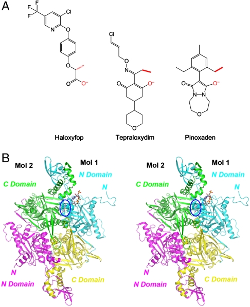
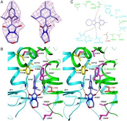
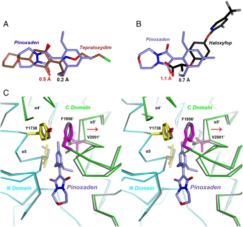
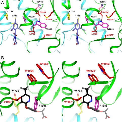
Acetyl-CoA carboxylases (ACCs) are crucial metabolic enzymes and have been targeted for drug development against obesity, diabetes, and other diseases. The carboxyltransferase (CT) domain of this enzyme is the site of action for three different classes of herbicides, as represented by haloxyfop, tepraloxydim, and pinoxaden. Our earlier studies have demonstrated that haloxyfop and tepraloxydim bind in the CT active site at the interface of its dimer. However, the two compounds probe distinct regions of the dimer interface, sharing primarily only two common anchoring points of interaction with the enzyme. We report here the crystal structure of the CT domain of yeast ACC in complex with pinoxaden at 2.8-Å resolution. Despite their chemical diversity, pinoxaden has a similar binding mode as tepraloxydim and requires a small conformational change in the dimer interface for binding. Crystal structures of the CT domain in complex with all three classes of herbicides confirm the importance of the two anchoring points for herbicide binding. The structures also provide a foundation for understanding the molecular basis of the herbicide resistance mutations and cross resistance among the herbicides, as well as for the design and development of new inhibitors against plant and human ACCs.


Conflict of interest statement

The authors declare no conflict of interest.

Figures

Fig. 1.
Fig. 1.
Crystal structure of the CT domain in complex with pinoxaden. (A) Chemical structures of the herbicides haloxyfop, tepraloxydim, and pinoxaden. The two anchoring points for interactions with the CT domain (see text for details) are colored in red. (B) Schematic drawing of the structure of yeast CT domain dimer in complex with pinoxaden. The N domains of the two monomers are colored in cyan and magenta, while the C domains are colored in yellow and green. The inhibitor is shown in stick models, in slate blue for carbon atoms, and one inhibitor molecule is highlighted by the blue oval. The CoA molecule is shown for reference in gray (13). The structure figures were produced with PyMOL (www.pymol.org).
Fig. 2.
Fig. 2.
The binding mode of pinoxaden. (A) Final omit Fo-Fc electron density at 2.8-Å resolution for pinoxaden, contoured at 3σ, in two views. (B) Stereographic drawing showing the binding site for pinoxaden. The N domain of one monomer is colored in cyan, and the C domain of the other monomer in green. The side chains of residues in the binding site are shown in yellow and magenta, respectively. Leu1705 is shown and labeled in red, because it is equivalent to a site of resistance mutations in plants. Hydrogen bonds from the inhibitor to the protein are indicated with dashed red lines. (C) Schematic drawing of the interactions between pinoxaden and the CT active site.
Fig. 3.
Fig. 3.
Comparison of the binding modes of pinoxaden with haloxyfop and tepraloxydim. (A) Overlay of the binding modes of pinoxaden (in slate blue for carbon atoms) and tepraloxydim (brown). The distances between the oxyanions and methyl groups of the two inhibitors are indicated. (B) Overlay of the binding modes of pinoxaden and haloxyfop (black). (C) Structural overlay of the CT domain free enzyme (in gray) and the pinoxaden complex (in cyan and green for the N and C domains) near the inhibitor binding site. Side chains in the binding site with large conformational changes are also shown. Phe1956′ is shown for reference. The shift in the position of the α5′ helix is indicated with the red arrow.
Fig. 4.
Fig. 4.
Molecular basis for herbicide resistance mutations. (A) Stereographic drawing showing the structure of yeast CT domain near the mutation sites I1781L (L1705 in yeast ACC), D2078G (D2004′), C2088R (M2014′), and G2096A (A2022′). The bound position of pinoxaden is also shown. (B) Stereographic drawing showing the structure of yeast CT domain near the mutation sites W1999C (W1924′ in yeast ACC), W2027C (W1953′), and I2041N (V1967′). The bound position of haloxyfop is also shown.

References

    1. Wakil SJ, Stoops JK, Joshi VC. Fatty acid synthesis and its regulation. Annu Rev Biochem. 1983;52:537–579. - PubMed
    1. Tong L. Acetyl-coenzyme A carboxylase: Crucial metabolic enzyme and attractive target for drug discovery. Cell Mol Life Sci. 2005;62:1784–1803. - PMC - PubMed
    1. McGarry JD, Brown NF. The mitochondrial carnitine palmitoyltransferase system. From concept to molecular analysis. Eur J Biochem. 1997;244:1–14. - PubMed
    1. Ramsay RR, Gandour RD, van der Leij FR. Molecular enzymology of carnitine transfer and transport. Biochim Biophys Acta. 2001;1546:21–43. - PubMed
    1. Abu-Elheiga L, Matzuk MM, Abo-Hashema KAH, Wakil SJ. Continuous fatty acid oxidation and reduced fat storage in mice lacking acetyl-CoA carboxylase 2. Science. 2001;291:2613–2616. - PubMed

Publication types

MeSH terms

Associated data

LinkOut - more resources