. 2010 Dec 21;107(51):22084-9.
doi: 10.1073/pnas.1012498107. Epub 2010 Dec 6.

Opn5 is a UV-sensitive bistable pigment that couples with Gi subtype of G protein

Affiliations

Opn5 is a UV-sensitive bistable pigment that couples with Gi subtype of G protein

Takahiro Yamashita et al. Proc Natl Acad Sci U S A. .

Abstract

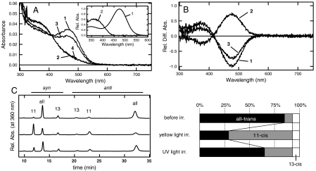
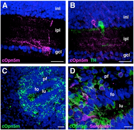
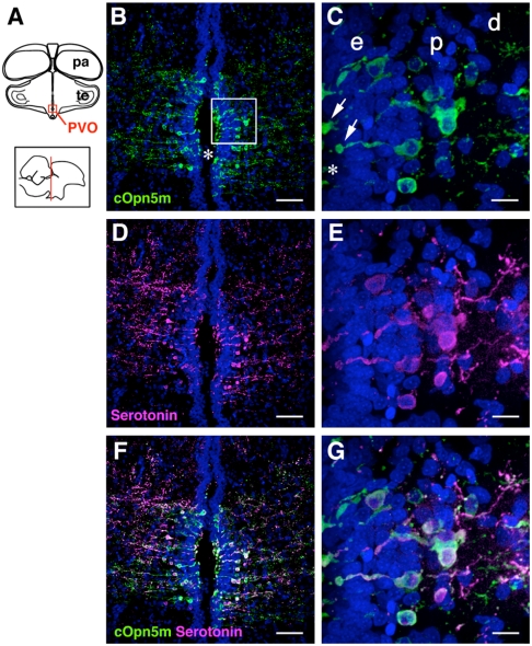
Opn5 (neuropsin) belongs to an independent group separated from the other six groups in the phylogenetic tree of opsins, for which little information of absorption characteristics and molecular properties of the members is available. Here we show that the chicken Opn5 (cOpn5m) is a UV-sensitive bistable pigment that couples with Gi subtype of G protein. The recombinant expression of cOpn5m in HEK 293s cells followed by the addition of 11-cis- and all-trans-retinal produced UV light-absorbing and visible light-absorbing forms, respectively. These forms were interconvertible by UV and visible light irradiations, respectively, indicating that cOpn5m is a bistable pigment. The absorption maxima of these forms were estimated to be 360 and 474 nm, respectively. The GTPγS binding assay clearly showed that the visible light-absorbing form having all-trans-retinal activates Gi type of G protein, whereas no Gt or Gq activation ability was observed. Immunohistochemical studies using an antibody against cOpn5m clearly showed that this pigment is localized within some types of amacrine cells and some cells in the ganglion cell layer of the retinas, the vast majority of cells in the pineal gland and serotonin-positive cells in the paraventricular organ. Because cOpn5m is the only UV-sensitive opsin among the opsins found so far in chicken, this study provides the molecular basis for UV reception in chicken.


Conflict of interest statement

The authors declare no conflict of interest.

Figures

Fig. 1.
Fig. 1.
Absorption spectra and retinal configurations of cOpn5m. (A) Absorption spectra of purified cOpn5m reconstituted with all-trans-retinal. Spectra were measured in the dark (curve 1), after yellow light (> 500 nm) irradiation (curve 2), and after subsequent UV light irradiation (curve 3), and reirradiation with yellow light (curve 4). (Inset) The calculated absorption spectra of cOpn5m in the dark (curve 1) and after yellow light irradiation (curve 2). The calculation procedures are described in the text. (B) Spectral change caused by irradiation with yellow light (curve 1) and subsequent UV light (curve 2) and reirradiation with yellow light (curve 3). (C) Retinal configuration changes by yellow light and subsequent UV light irradiations of cOpn5m. (Left) The retinal configurations were analyzed with HPLC after extraction of the chromophore as retinal oximes (syn and anti forms of 11-cis, 13-cis, and all-trans retinal oximes). (Right) Isomeric compositions of retinal before and after light irradiations of cOpn5m.
Fig. 2.
Fig. 2.
Time courses of G protein activation ability by cOpn5m. Gi (A), Gt (B), and Gq (C) activation abilities of cOpn5m reconstituted with 11-cis-retinal were measured in the dark (open circle), after yellow light (> 500 nm) irradiation (open square), and after subsequent UV light irradiation (open triangle). Experiments were performed at 0 °C. Data are presented as the means ± SD of three independent experiments.
Fig. 3.
Fig. 3.
cOpn5m photoreceptors in the posthatching chick retina (P14) and pineal gland (P11). (A) A cOpn5m-immunoreactive (IR) cell (magenta) in the ganglion cell layer of retina. gcl, ganglion cell layer; ipl, inner plexiform layer; inl, inner nuclear layer. (B) A cOpn5m-IR cells (magenta) in the vicinity of tyrosine hydroxylase (TH)-IR cells (green). (C) cOpn5m-IR cells (green) are also present in the pineal gland. fo, follicular cells; lu, follicular lumen; pf, parafollicular cells. (D) Enlargement of the pineal gland in C. cOpn5m-immunoreactivity is found in the outer segments of the pinealocytes facing the lumen and perikarya of parafollicular cells. cOpn5m-positive outer segments of pineal photoreceptors are mostly serotonin-positive (magenta). (Scale bars: 10 μm, D; 25 μm, AC.)
Fig. 4.
Fig. 4.
cOpn5m photoreceptors in the posthatching chick deep brain (P11). (A) Schematic diagram of chick brain. (Upper) Coronal plane; (Lower) sagittal plane. Boxed region is shown in B, D, and F. pa, pallium; PVO, paraventricular organ; te, tectum. (B) cOpn5m-IR cells (green) are predominantly present in the chicken PVO, where serotonin-IR cells (magenta) are mainly located (D) (46). Asterisk, the third ventricle. Nuclei of the cells are stained with DAPI (blue). The boxed region in B is enlarged in C, E, and G. (C) cOpn5m-IR cells are bipolar and their club-like projections (arrows) line the third ventricle. They are present in the ependymal layer (e) and proximal part (p) of the PVO. d, distal part of the nucleus of the PVO (16). (F and G) Most of the Opn5m-IR cells in the PVO are overlapping with serotonin-IR cells. (Scale bars: 10 μm, C, E, and G; 50 μm, B, D, and F.)

References

    1. Koyanagi M, Terakita A. Gq-coupled rhodopsin subfamily composed of invertebrate visual pigment and melanopsin. Photochem Photobiol. 2008;84:1024–1030. - PubMed
    1. Shichida Y, Matsuyama T. Evolution of opsins and phototransduction. Philos Trans R Soc London B. 2009;364:2881–2895. - PMC - PubMed
    1. Hofmann KP, et al. A G protein-coupled receptor at work: The rhodopsin model. Trends Biochem Sci. 2009;34:540–552. - PubMed
    1. Yau KW, Hardie RC. Phototransduction motifs and variations. Cell. 2009;139:246–264. - PMC - PubMed
    1. Shen D, et al. A human opsin-related gene that encodes a retinaldehyde-binding protein. Biochemistry. 1994;33:13117–13125. - PubMed

Publication types

MeSH terms

Substances