. 2011 Jan;9(1):115-30.
doi: 10.1158/1541-7786.MCR-10-0105. Epub 2010 Dec 6.

AP-1 regulates cyclin D1 and c-MYC transcription in an AKT-dependent manner in response to mTOR inhibition: role of AIP4/Itch-mediated JUNB degradation

Affiliations

AP-1 regulates cyclin D1 and c-MYC transcription in an AKT-dependent manner in response to mTOR inhibition: role of AIP4/Itch-mediated JUNB degradation

Raffi Vartanian et al. Mol Cancer Res. 2011 Jan.

Abstract

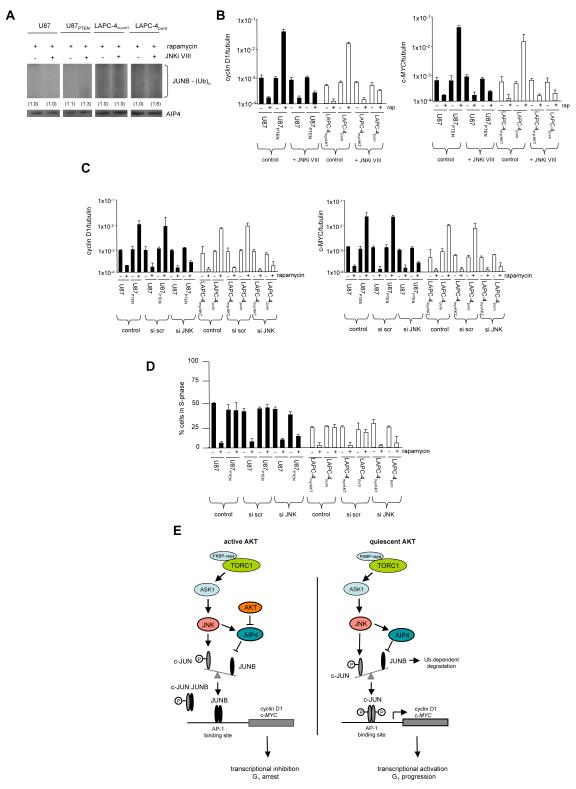
One mechanism by which AKT kinase-dependent hypersensitivity to mammalian target of rapamycin (mTOR) inhibitors is controlled is by the differential expression of cyclin D1 and c-MYC. Regulation of posttranscriptional processes has been demonstrated to be crucial in governing expression of these determinants in response to rapamycin. Our previous data suggested that cyclin D1 and c-MYC expression might additionally be coordinately regulated in an AKT-dependent manner at the level of transcription. Under conditions of relatively quiescent AKT activity, treatment of cells with rapamycin resulted in upregulation of cyclin D1 and c-MYC nascent transcription, whereas in cells containing active AKT, exposure repressed transcription. Promoter analysis identified AKT-dependent rapamycin responsive elements containing AP-1 transactivation sites. Phosphorylated c-JUN binding to these promoters correlated with activation of transcription whereas JUNB occupancy was associated with promoter repression. Forced overexpression of JunB or a conditionally active JunB-ER allele repressed cyclin D1 and c-MYC promoter activity in quiescent AKT-containing cells following rapamycin exposure. AIP4/Itch-dependent JUNB protein degradation was found to be markedly reduced in active AKT-containing cells compared with cells harboring quiescent AKT. Moreover, silencing AIP4/Itch expression or inhibiting JNK mediated AIP4 activity abrogated the rapamycin-induced effects on cyclin D1 and c-MYC promoter activities. Our findings support a role for the AKT-dependent regulation of AIP4/Itch activity in mediating the differential cyclin D1 and c-MYC transcriptional responses to rapamycin.


Figures

Figure 1
Figure 1
AKT-dependent alterations in cyclin D1 and c-MYC promoter activities following rapamycin exposure. (A) Cyclin D1 and (B) c-MYC AKT-dependent RNA polymerase II (Pol II) association following rapamycin treatment. The indicated cell lines were treated without or with rapamycin and Pol II occupancy determined. ChIP-quantitative PCR data are expressed as a ratio of cyclin D1 or c-MYC to tubulin. Mean + S.D. are shown, n = 3. (C) Nascent transcription of cyclin D1 and c-MYC mRNAs from the indicated lines treated without or with rapamycin. Actin and pBlueScript KS probes are also shown. Band intensities were quantified by densitometry and fold differences relative to non-treated control values are shown in parentheses below. (D) Relative levels of the indicated proteins from the indicated cell lines treated without or with rapamycin. (E) AKT-dependent effects on cell cycle distributions of the indicated cell lines following rapamycin exposure. Mean + S.D. are shown, n = 3.
Figure 2
Figure 2
Deletion analysis of the cyclin D1 and c-MYC promoters. The indicated constructs were transfected into U87 or U87PTEN cells, treated without or with rapamycin and luciferase activities determined. The indicated AP-1 sites within the constructs −1749wt and −2052HBMwt were mutated (shown in red) as described in materials and methods. The relative fold change in luciferase activity is shown as compared to untreated controls. Mean + S.D. are shown, n = 3.
Figure 3
Figure 3
AKT-dependent c-JUN and JUNB differential binding to the cyclin D1 and c-MYC promoters following rapamycin treatment. (A) EMSA analysis using 32P-labeled cyclin D1 (left panel) or c-MYC (right panel) DNA probes containing native AP-1 binding sites were carried out using the indicated nuclear extracts in the absence or presence of control (IgG) or antibody against c-JUN. (B) as in (A) except antibody to JUNB was used. Arrows indicate super-shifted species. Experiments were carried out three times with similar results. (C) Extracts from U87 and U87PTEN cells untreated or treated with rapamycin were chromatin immunoprecipitated with the indicated antibodies (c-JUN, top panels; JUNB, bottom panels) and fragments were subjected to quantitative real-time PCR analysis using various primer sets spanning the promoter regions (cyclin D1, left panels; c-MYC, right panels) as indicated by the gray bars. Primer sets 1 correspond to the AP-1 binding site(s) in the cyclin D1 and c-MYC promoters. At least two independent experiments were performed and the mean + S.D. are shown. (D) Sepharose beads were conjugated with cyclin D1 promoter element DNA (D1-beads, left panel) or c-MYC promoter element (MYC-beads, right panel) containing the native AP-1 binding site(s) or beads without linked DNA were incubated with nuclear extracts from U87 or U87PTEN cells treated without or with rapamycin as indicated. Following recovery by centrifugation and washing of the beads, bound material was analyzed by immunoblot for the indicated proteins. These experiments were performed twice with similar results.
Figure 4
Figure 4
Modulation of JUNB regulates AKT-dependent cyclin D1 and c-MYC promoter activity and rapamycin sensitivity. (A) Control or JUNB overexpressing cells (U87 and U87PTEN, shaded bars; LAPC-4myrAKT and LAPC-4puro, open bars) were treated without or with rapamycin and Pol II promoter occupancy determined for cyclin D1 (left panel) and c-MYC (right panel) as in Fig. 1. Mean + S.D. are shown, n = 3. (B) Effects of rapamycin on Pol II promoter occupancy of cyclin D1 (left panel) or c-MYC (right panel) in the indicated lines (U87 and U87PTEN, shaded bars; LAPC-4myrAKT and LAPC-4puro, open bars) expressing the JunB-ER fusion in the absence or presence of 4OHT (1 mM). Mean + S.D. are shown, n = 3. (C) Effects of JUNB knockdown on rapamycin-induced alterations in cyclin D1 (left panel) and c-MYC (right panel) Pol II promoter activity in the indicated cell lines. Mean + S.D. are shown, n = 3. (D) S-phase cell cycle analysis of JUNB overexpressing U87 and LAPC-4 cell line pairs following rapamycin exposure. Mean + S.D., n = 3. (E) Cell cycle analysis of JUNB siRNA-treated U87 and LAPC-4 cell line pairs following rapamycin exposure. Mean + S.D., n = 3.
Figure 5
Figure 5
AKT-dependent variations in JUNB protein ubiquitination, AIP4 protein levels and activity following rapamycin exposure. (A) JUNB ubiquitination in U87, U87PTEN, LAPC-4myrAKT and LAPC-4puro cells following rapamycin exposure as indicated. Cells were transfected with an HA-ubiquitin construct, treated without or with rapamycin and extracts immunoprecipitated using JUNB antibodies and immunoprecipitates subsequently immunoblotted for the extent of HA-ubiquitination using α-HA antibody and total JUNB. Relative densitometric values are shown in parentheses below. (B) Relative AIP4, JUN and actin protein levels following rapamycin exposure (10 nM) for various time points from the indicated cell lines. (C) AIP4 activity in U87, U87PTEN, LAPC-4myrAKT and LAPC-4puro cells following rapamycin exposure. Cells were treated with rapamycin as indicated and AIP4 immunoprecipitates utilized in in vitro ubiquitination reactions using GST-JUNB as a substrate. Poly-ubiquinated JUNB was visualized by immunoblotting using α-JUNB antibodies.
Figure 6
Figure 6
Knockdown of AIP4 abrogates AKT-dependent cyclin D1 and c-MYC promoter activity following mTOR inhibition. (A) U87, U87PTEN, LAPC-4myrAKT or LAPC-4puro cells were treated with siRNA targeting AIP4 or a scrambled (scr) nontargeting control siRNA in the presence of rapamycin and extracts immunoblotted for the indicated proteins. (B) Cyclin D1 (left panel) and c-MYC (right panel) promoter Pol II occupancy in the paired cell lines (U87 and U87PTEN, shaded bars; LAPC-4myrAKT and LAPC-4puro, open bars) following treatment with the indicated siRNAs and rapamycin. Mean + S.D. are shown, n = 3. (C) S-phase cell cycle analysis of AIP4 siRNA-treated treated cell lines in response to rapamycin exposure. Mean + S.D., n =3.
Figure 7
Figure 7
JNK activity is required for differential AKT-dependent AIP4 activity, cyclin D1 and c-MYC transcriptional responses and G1 arrest in response to rapamycin. (A) Effects of JNK inhibitor (JNKi VIII) on in vitro AIP4 activity. The paired cell lines were treated with rapamycin and JNKi VIII (4 μM) as indicated and AIP4 immune-complexes used in in vitro ubiquitination reactions as in Fig. 5 (C). (B) Cyclin D1 (left panel) and c-MYC (right panel) Pol II association in the cell lines (U87 and U87PTEN, shaded bars; LAPC-4myrAKT and LAPC-4puro, open bars) following treatments with JNKi VIII (4 μM) and rapamycin as indicated. Mean + S.D. are shown, n = 3. (C) Cyclin D1 and c-MYC Pol II occupancy in U87, U87PTEN, LAPC-4myrAKT and LAPC-4puro cells treated with nontargeting scrambled (scr) or JNK siRNA as indicated in the presence of rapamycin (U87 and U87PTEN, shaded bars; LAPC-4myrAKT and LAPC-4puro, open bars). Mean + S.D. are shown, n = 3. (D) S-phase cell cycle analysis of paired cell lines treated with nontargeting (scr) or JNK siRNAs in the presence of rapamycin. Mean + S.D., n = 3. (E). Regulation of cyclin D1 and c-MYC transcription by AP-1 in response to rapamycin. In this model, rapamycin-FKBP12 inactivation of TORC1 results in sustained activation of the JNK cascade via increased ASK1 activity (27, 52). This leads to AIP4-mediated JUNB ubiquitination and degradation in quiescent AKT-containing cells and is inhibited in tumor cells whose AKT activity is elevated. As a result, accumulated JUNB and c-JUN-JUNB heterodimers bind to AP-1 elements within the cyclin D1 and c-MYC promoters and inhibits transcription. In contrast, in cells with relatively quiescent AKT, rapamycin exposure results in activation of JNK/AIP4 signaling, degradation of JUNB and stimulation of cyclin D1 and c-MYC promoters via c-JUN.

References

    1. Guertin DA, Sabatini DM. The pharmacology of mTOR inhibition. Sci Signal. 2009;2:pe24. - PubMed
    1. Guertin DA, Sabatini DM. Defining the role of mTOR in cancer. Cancer Cell. 2007;12:9–22. - PubMed
    1. Easton JB, Houghton PJ. mTOR and cancer therapy. Oncogene. 2006;25:6436–46. - PubMed
    1. Sabatini DM. mTOR and cancer: insights into a complex relationship. Nat Rev Cancer. 2006;6:729–34. - PubMed
    1. Proud CG. mTORC1 signalling and mRNA translation. Biochem Soc Trans. 2009;37:227–31. - PubMed

Publication types

MeSH terms