. 2011 Feb 10;117(6):1928-37.
doi: 10.1182/blood-2010-09-305649. Epub 2010 Dec 6.

The novel receptor tyrosine kinase Axl is constitutively active in B-cell chronic lymphocytic leukemia and acts as a docking site of nonreceptor kinases: implications for therapy

Affiliations

The novel receptor tyrosine kinase Axl is constitutively active in B-cell chronic lymphocytic leukemia and acts as a docking site of nonreceptor kinases: implications for therapy

Asish K Ghosh et al. Blood. .

Abstract

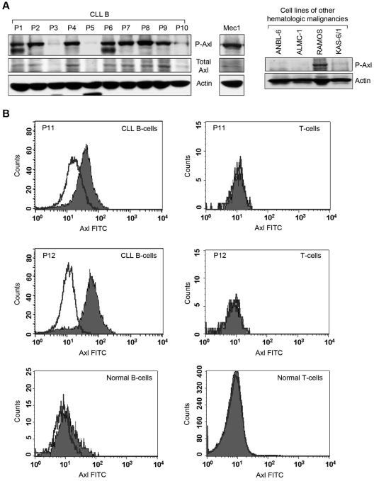
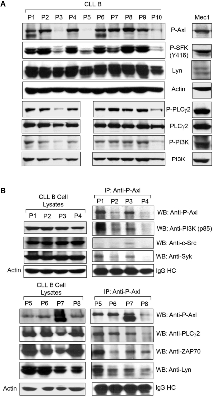
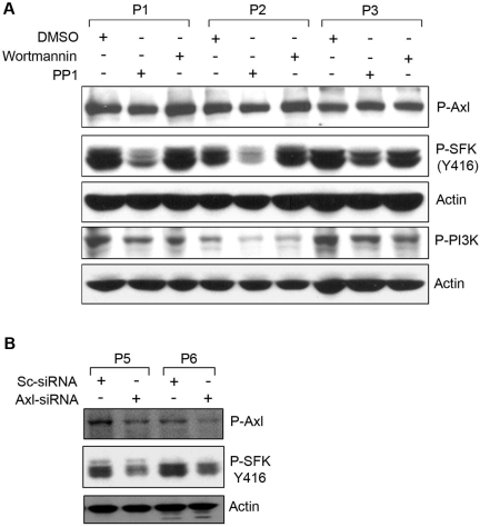
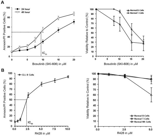
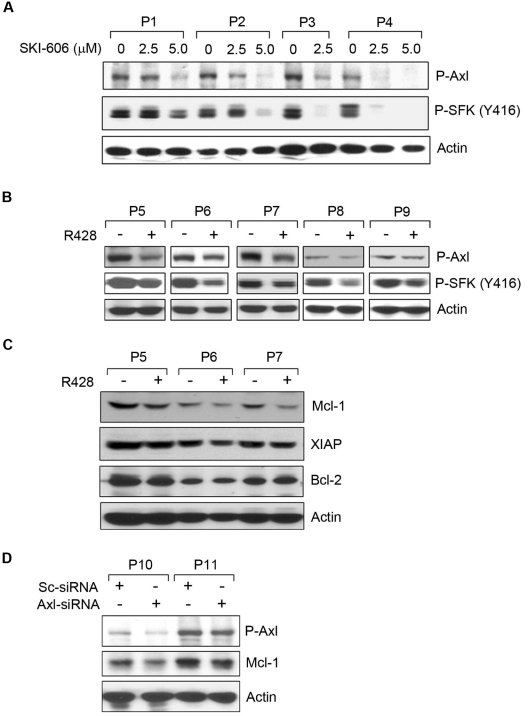
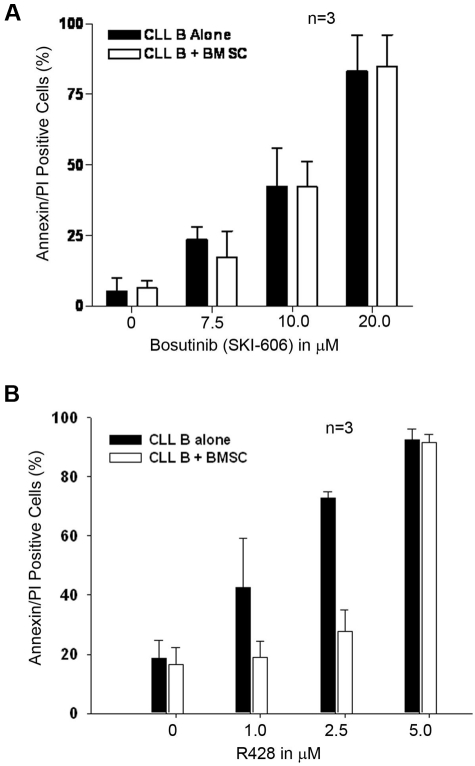
Recently, we detected that chronic lymphocytic leukemia (CLL) B-cell-derived microvesicles in CLL plasma carry a constitutively phosphorylated novel receptor tyrosine kinase (RTK), Axl, indicating that Axl was acquired from the leukemic B cells. To examine Axl status in CLL, we determined the expression of phosphorylated-Axl (P-Axl) in freshly isolated CLL B cells by Western blot analysis. We detected differential levels of P-Axl in CLL B cells, and further analysis showed that expression of P-Axl was correlated with the other constitutively phosphorylated kinases, including Lyn, phosphoinositide-3 kinase, SyK/ζ-associated protein of 70 kDa, phospholipase C γ2 in CLL B cells. We found that these intracellular signaling molecules were complexed with P-Axl in primary CLL B cells. When Axl and Src kinases were targeted by a Src/Abl kinase inhibitor, bosutinib (SKI-606), or a specific-inhibitor of Axl (R428), robust induction of CLL B-cell apoptosis was observed in both a dose- and time-dependent manner. Therefore, we have identified a novel RTK in CLL B cells which appears to work as a docking site for multiple non-RTKs and drives leukemic cell survival signals. These findings highlight a unique target for CLL treatment.


Figures

Figure 1
Figure 1
Primary CLL B cells express constitutively P-Axl. (A) Detection of Axl in primary CLL B cells and other hematologic malignancies. Lysates of CLL B cells from patients with CLL (n = 10) and Mec-1 cell line were examined for the presence of P-Axl by Western blot analysis. Expression of total Axl was also examined. Lysates of 3 myeloma cell lines (ANBL-6, ALMC-1, and KAS-6/1) and 1 cell line from Burkitt lymphoma (RAMOS) were also examined for the detection of phospho-Axl in Western blot analysis. Individual patients (P) are indicated and sequentially numbered. Actin was used as loading control. (B) Detection of cell surface Axl. Peripheral blood mononuclear cells from patients with CLL or healthy persons were stained with mouse monoclonal antibodies to CD19 and CD5 conjugated with allophycocyanin or phycoerythrin, respectively, and an unconjugated antibody to human Axl, followed by FITC-conjugated secondary antibody or respective fluorescein-conjugated isotype controls. Cells were washed and analyzed for the expression of Axl by gating CD19+/CD5+ (CLL B cell), CD19+/CD5 (normal B cell) or CD19/CD5+ (T cell) populations. Histogram overlay diagram of 2 representative patients with CLL (P11 and P12) and 1 healthy control are shown. Black line indicates isotype controls and filled histogram indicates Axl expression.
Figure 2
Figure 2
P-Axl is coexpressed with multiple constitutively phosphorylated non-RTKs and serving as a docking site in CLL B cells. (A) Expression of P-Axl is associated with coexpression of various phospho-nonreceptor kinases. CLL B-cell lysates from the same patient cohort as in Figure 1A were examined for the expression of various phosphorylated intracellular kinases shown by Western blot analysis. Actin was used as loading control. (B) P-Axl is physically associated with multiple nonreceptor kinases in CLL B cells. P-Axl was immunoprecipitated from purified CLL B-cell lysates from patients with CLL (n = 8) using a phospho-Axl–specific antibody. The precipitated immunocomplex was examined by Western blot analysis for the presence of various kinases. The IgG heavy chain (IgG HC) was shown for equal loading of the immunocomplex. Expression levels of these kinases in CLL B-cell lysates were also examined (left) for comparison. Actin was used as loading control for the cell lysates. Patients with CLL were assigned arbitrary numbers and are different from the cohort in Figure 1.
Figure 3
Figure 3
Phosphorylation of Axl is an upstream event of SFK activation. (A) Inhibition of SFK phosphorylation does not alter Axl phosphorylation. Cell lysates from CLL B cells treated with the Src inhibitor (PP1) or PI3K inhibitor (wortmannin) or left untreated were examined for the detection of phosphorylated Axl, SFK, or PI3K by Western blot analysis. Actin was used as loading control. (B) Knockdown of Axl reduces phosphorylation of SFK. CLL B cells were transfected with scrambled siRNA (Sc-siRNA) as a control or Axl-specific siRNA, and cell lysates were analyzed for the detection of phosphorylated Axl and SFK in Western blot. Actin was used as loading control. Patients with CLL are indicated by arbitrary numbers.
Figure 4
Figure 4
Targeting Axl and SFK induces apoptosis in CLL B cells. (A) Bosutinib (SKI-606) induces apoptosis in CLL B cells. Freshly isolated CLL B cells (n = 20) were treated with increasing doses of SKI-606 as indicated for 24 and 48 hours. Cells were harvested and stained with Annexin/PI for flow cytometric analysis. Mean values are presented with standard error bars. Effect of SKI-606 on normal B and T lymphocytes (n = 3) were also examined after a 24-hour incubation with the indicated doses of SKI-606. (B) Specific Axl inhibitor R428 induces apoptosis in CLL B cells. Freshly isolated CLL B cells (n = 10) were treated with increasing doses of R428 as indicated for 24 hours. Cells were stained with Annexin/PI for flow cytometric analysis as described. Mean values are presented with standard error bars. Effect of R428 on normal B and T lymphocytes and NK cells (n = 4) were also examined similarly after a 24-hour incubation with the indicated doses of R428.
Figure 5
Figure 5
Induction of apoptosis in CLL B cells by SKI-606 or R428 is associated with reduction of Axl phosphorylation. (A) SKI-606 targets Axl and SFK phosphorylation. Primary CLL B cells were treated with SKI-606 at sublethal doses as indicated for 24 hours. Cell lysates were examined for levels of Axl and SFK (Tyr 416) phosphorylation by Western blot analysis. Actin was used as loading control. (B) R428 inhibits Axl phosphorylation. CLL B cells were treated with R428 at a suboptimal dose (based on apoptosis studies) of 1.0μM for 24 hours, and cell lysates were examined for the Axl and SFK phosphorylation by Western blot analysis with the use of specific antibodies. Actin was used as loading control. Patients with patients were indicated by assigning arbitrary numbers. (C) Axl inhibition reduces Mcl-1. R428-treated CLL B-cell lysates (P5-P7) used above (B) were analyzed for the expression of Mcl-1, XIAP, and Bcl-2 by Western blots with the use of specific antibodies. A consistent reduction of Mcl-1 was detected in these cell lysates; however; only a subtle, if at all, or no effects on the expression of XIAP or Bcl-2 was observed on Axl-inhibition. Actin was used as loading control. (D) Knockdown of Axl reduces Mcl-1. CLL B cells were transfected with scrambled siRNA (Sc-siRNA) as a control or Axl-specific siRNA, and cell lysates were analyzed after 48 hours of transfection for the detection of P-Axl and Mcl-1 in Western blot. Actin was used as loading control. Patients with CLL are indicated by arbitrary numbers.
Figure 6
Figure 6
Effect of SKI-606 and R428 on stroma-mediated protection of CLL B cells. (A) SKI-606 is able to overcome stromal-mediated protection. Freshly isolated CLL B cells (n = 3) were cocultured with the primary CLL BMSCs or cultured alone and treated with increasing doses of SKI-606 for 24 hours. Cells were harvested and stained with CD19 antibody and Annexin/PI for flow cytometric analysis. Mean values are presented with standard error bars. (B) Coculture of CLL B cells with CLL BMSCs protects from R428-induced apoptosis. Freshly isolated CLL B cells (n = 3) were cocultured with the primary CLL BMSCs or cultured alone and treated with increasing doses of R428 for 24 hours. CLL B cells were harvested and stained with CD19 antibody and Annexin/PI for flow cytometric analysis. Mean values are presented with standard error bars.

References

    1. Pleyer L, Egle A, Hartmann TN, Greil R. Molecular and cellular mechanisms of CLL: novel therapeutic approaches. Nat Rev Clin Oncol. 2009;6(7):405–418. - PubMed
    1. Caligaris-Cappio F. Biology of chronic lymphocytic leukemia. Rev Clin Exp Hematol. 2000;4(1):5–21. - PubMed
    1. Hanahan D, Weinberg RA. The hallmarks of cancer. Cell. 2000;100(1):57–70. - PubMed
    1. Blume-Jensen P, Hunter T. Oncogenic kinase signalling. Nature. 2001;411(6835):355–365. - PubMed
    1. Zhang YX, Knyazev PG, Cheburkin YV, et al. AXL is a potential target for therapeutic intervention in breast cancer progression. Cancer Res. 2008;68(6):1905–1915. - PubMed

Publication types

MeSH terms