. 2011 Jan;121(1):238-48.
doi: 10.1172/JCI42520. Epub 2010 Dec 6.

Staphylococcus epidermidis surfactant peptides promote biofilm maturation and dissemination of biofilm-associated infection in mice

Affiliations

Staphylococcus epidermidis surfactant peptides promote biofilm maturation and dissemination of biofilm-associated infection in mice

Rong Wang et al. J Clin Invest. 2011 Jan.

Abstract

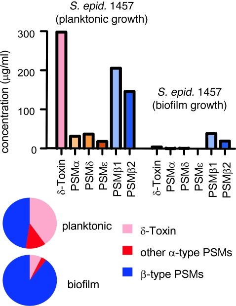
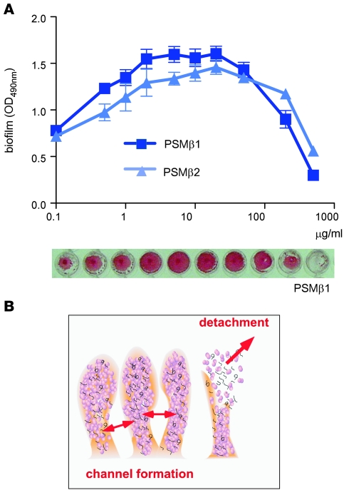
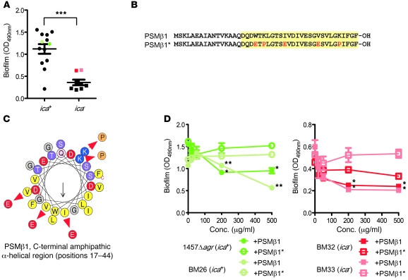
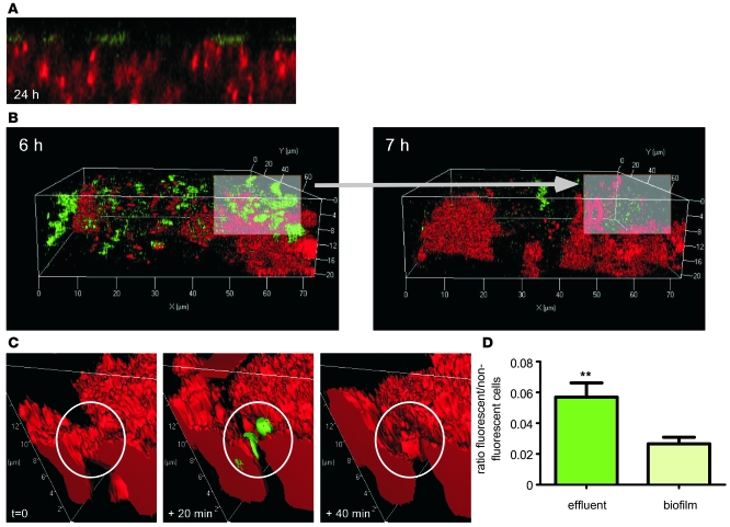
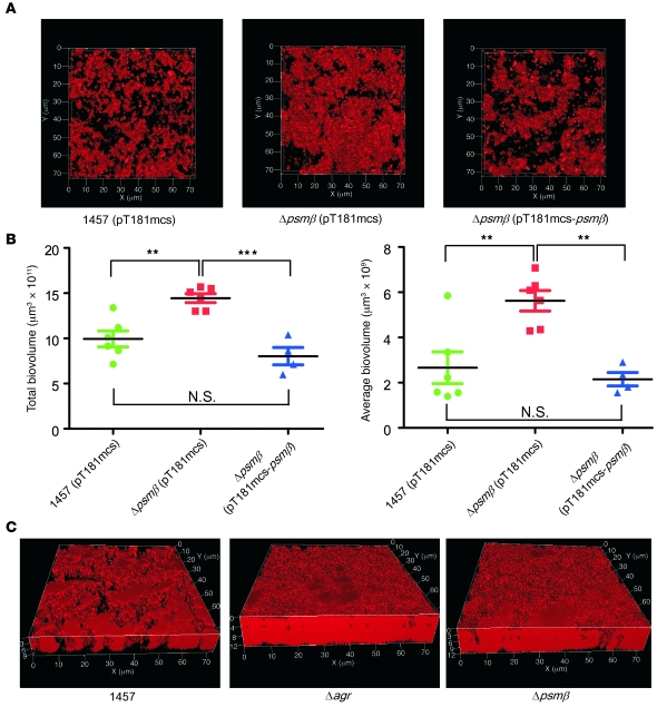
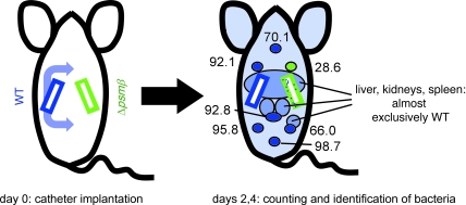
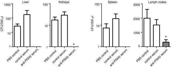
Biofilms are surface-attached agglomerations of microorganisms embedded in an extracellular matrix. Biofilm-associated infections are difficult to eradicate and represent a significant reservoir for disseminating and recurring serious infections. Infections involving biofilms frequently develop on indwelling medical devices in hospitalized patients, and Staphylococcus epidermidis is the leading cause of infection in this setting. However, the molecular determinants of biofilm dissemination are unknown. Here we have demonstrated that specific secreted, surfactant-like S. epidermidis peptides--the β subclass of phenol-soluble modulins (PSMs)--promote S. epidermidis biofilm structuring and detachment in vitro and dissemination from colonized catheters in a mouse model of device-related infection. Our study establishes in vivo significance of biofilm detachment mechanisms for the systemic spread of biofilm-associated infection and identifies the effectors of biofilm maturation and detachment in a premier biofilm-forming pathogen. Furthermore, by demonstrating that antibodies against PSMβ peptides inhibited bacterial spread from indwelling medical devices, we have provided proof of principle that interfering with biofilm detachment mechanisms may prevent dissemination of biofilm-associated infection.


Figures

Figure 1
Figure 1. PSM production under biofilm and planktonic modes of growth.
PSM production under planktonic (shaken 125 ml flasks containing 50 ml TSB/0.5% glucose, 18 hour growth, 37°C) or biofilm (24 hour static culture in microtiter plates, 37°C) modes of growth was assayed by RP-HPLC/ESI-MS. PSMs were determined in supernatants of planktonic or biofilm cultures after centrifugation. Absolute amounts are shown at the top, relative composition at the bottom. S. epid. 1457, S. epidermidis 1457.
Figure 2
Figure 2. The psmβ operon.
(A) Organization of the psmβ locus in genome-sequenced S. epidermidis strains and the 1457 strain used in this study. Construction of the psmβ mutant allelic replacement strain is shown at the bottom. (B) Northern blot analysis of S. epidermidis 1457 RNA from different growth phases using a psmβ probe. The psmβ signal is marked by an arrow.
Figure 3
Figure 3. S. epidermidis in vitro biofilm formation under influence of PSMβ peptides.
(A) Biofilm formation in microtiter plates (24 hours, 37°C). PSMβ peptides at different concentrations were added at the time of inoculation with S. epidermidis agr (devoid of PSMs). Biofilms were made visible using safranin staining (see example for PSMβ1 at the bottom), and biofilm formation was measured using an ELISA reader. Error bars depict mean ± SEM. (B) Schematic presentation of biofilm cell-cell disruptive processes leading to channel formation and detachment.
Figure 4
Figure 4. Specific impact of PSMβ1 on in vitro biofilm detachment.
(A) Biofilm formation by functionally agr-negative (without PSM production) ica-positive versus ica-negative strains. Biofilms were grown in microtiter plates for 24 hours and stained with safranin. ***P < 0.0001, t test. Colored symbols represent the strains used for the analyses shown in D. Error bars depict mean ± SEM. (B) α-Helical wheel presentation and (C) sequences of PSMβ1 and the mutated PSMβ1* peptide. The α-helical part (positions 17–44) is shaded in yellow. In the sequence, amino acids changed in PSMβ1* in comparison with PSMβ1 are shown in red; in the wheel presentation, exchanges are shown using red arrows. (D) Impact of PSMβ1 versus PSMβ1* on in-vitro biofilm development by 2 ica-positive and 2 ica-negative S. epidermidis strains. Conditions are the same as those used for the experiment shown in Figure 3A. *P < 0.05; **P < 0.01, t tests comparing values for the PSMβ1- versus PSMβ1*-treated samples at corresponding concentrations of added peptide.
Figure 5
Figure 5. Role of PSMβ peptides in S. epidermidis biofilm development: cell detachment.
A psmβ-promoter egfp fusion construct (see Supplemental Figure 1 for the synthetic egfp gene with optimized Staphylococcus codon usage) was produced to monitor psmβ expression in dynamic S. epidermidis biofilms using flow cells with CLSM (green). Entire biofilms were stained with propidium iodide (red). (A) Mature (24 hours) biofilm showing expression of psmβ at the outer biofilm layer. (B and C) Biofilm cluster detachment observed during biofilm development. Lower (A) and higher (B) temporal and spatial resolution is shown. (D) Comparative analysis of the relative amount of cells expressing psmβ (green fluorescence) in effluent and biofilm. The ratio of fluorescent versus nonfluorescent cells was determined using IMARIS software in effluent and biofilm samples. **P < 0.01, t test. Error bars depict mean ± SEM.
Figure 6
Figure 6. Role of PSMβ peptides and agr in S. epidermidis biofilm development: channel formation and biofilm expansion.
(A) CLSM pictures of S. epidermidis 1457 WT, isogenic psmβ mutant, and psmβ-complemented strains. The WT and psmβ mutant strains were transformed with the control plasmid pT181mcs to ensure comparability. Static biofilms were grown for 24 hours, stained with propidium iodide, and imaged using CLSM. View is from the top. (B) Analyses of total and average biovolumes, which are measures of total biofilm and degree of channel formation, respectively, using IMARIS software of the biofilm samples shown in part A. Note that increased average biofilm volume corresponds to decreased channel formation. **P < 0.01; ***P < 0.001, 1-way ANOVA with Bonferroni’s post tests. Error bars depict mean ± SEM. (C) CLSM pictures of S. epidermidis 1457 WT, isogenic agr, and psmβ operon deletion mutant biofilms. Growth conditions are as in A.
Figure 7
Figure 7. Role of PSMβ peptides in the dissemination of biofilm-associated infection.
Catheter pieces with equal cell numbers of preformed biofilms of either the S. epidermidis 1457 WT or isogenic psmβ mutant strains were inserted under the skin of mice at the dorsum. Each mouse received 2 pieces, left and right, 1 of which was coated with WT and 1 with mutant biofilms. At days 2 and 4 after insertion (this timing found in a pilot experiment to be optimal), catheter pieces were removed and dissemination was analyzed. In a first experiment, body fluids (from the i.p. cavity) and organs were analyzed. Almost all disseminated bacteria detected were of the WT, and significantly more WT than mutant bacteria were detected in the body fluids and liver. Number of mice: n = 9 (day 2); n = 6 (day 4). In a second experiment, lymph nodes at day 2 after infection were analyzed and showed predominantly WT bacteria, except for in the nodes adjacent to the catheter infected with psmβ mutant bacteria. Numbers represent percentages of WT among total bacteria from all tested mice (n = 7) in the respective lymph nodes. Blue, WT; green, psmβ mutant strains. See Table 1 for details.
Figure 8
Figure 8. Blocking dissemination of biofilm-associated infection using anti-PSMβ serum.
Specific antisera against PSMβ1 and PSMβ2 were mixed 1:1 and used to protect mice in the biofilm infection/dissemination model. *P < 0.05, versus PBS and control serum for the lymph nodes, versus PBS for the kidney results; 1-way ANOVA with Bonferroni’s post-tests. For spleen and liver, differences were not statistically significant owing to the fact that dissemination into organs only occurred in a limited number of mice and therefore SEM values were high. However, dissemination into the organs of mice that received anti-PSMβ serum was never observed (average CFU/100 μl ≤ 1). Number of mice: controls, n = 9; anti-PSMβ, n = 7. Control serum was from animals that received injections containing CFA and IFA. Error bars depict mean ± SEM.

Similar articles

Cited by

References

    1. Costerton JW, Stewart PS, Greenberg EP. Bacterial biofilms: a common cause of persistent infections. Science. 1999;284(5418):1318–1322. doi: 10.1126/science.284.5418.1318. - DOI - PubMed
    1. Mah TF, O’Toole GA. Mechanisms of biofilm resistance to antimicrobial agents. Trends Microbiol. 2001;9(1):34–39. doi: 10.1016/S0966-842X(00)01913-2. - DOI - PubMed
    1. Otto M. Staphylococcus epidermidis - the ‘accidental’ pathogen. . Nat Rev Microbiol. 2009;7(8):555–567. doi: 10.1038/nrmicro2182. - DOI - PMC - PubMed
    1. Rogers KL, Fey PD, Rupp ME. Coagulase-negative staphylococcal infections. Infect Dis Clin North Am. 2009;23(1):73–98. doi: 10.1016/j.idc.2008.10.001. - DOI - PubMed
    1. Stoll BJ, et al. Late-onset sepsis in very low birth weight neonates: the experience of the NICHD Neonatal Research Network. Pediatrics. 2002;110(2 pt 1):285–291. doi: 10.1542/peds.110.2.285. - DOI - PubMed

Publication types

MeSH terms