. 2011 Jan;121(1):318-27.
doi: 10.1172/JCI44723. Epub 2010 Dec 6.

Death effector domain-containing protein (DEDD) is required for uterine decidualization during early pregnancy in mice

Affiliations

Death effector domain-containing protein (DEDD) is required for uterine decidualization during early pregnancy in mice

Mayumi Mori et al. J Clin Invest. 2011 Jan.

Abstract

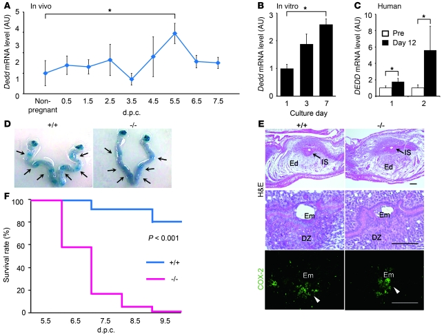
During intrauterine life, the mammalian embryo survives via its physical connection to the mother. The uterine decidua, which differentiates from stromal cells after implantation in a process known as decidualization, plays essential roles in supporting embryonic growth before establishment of the placenta. Here we show that female mice lacking death effector domain-containing protein (DEDD) are infertile owing to unsuccessful decidualization. In uteri of Dedd-/- mice, development of the decidual zone and the surrounding edema after embryonic implantation was defective. This was subsequently accompanied by disintegration of implantation site structure, leading to embryonic death before placentation. Polyploidization, a hallmark of mature decidual cells, was attenuated in DEDD-deficient cells during decidualization. Such inefficient decidualization appeared to be caused by decreased Akt levels, since polyploidization was restored in DEDD-deficient decidual cells by overexpression of Akt. In addition, we showed that DEDD associates with and stabilizes cyclin D3, an important element in polyploidization, and that overexpression of cyclin D3 in DEDD-deficient cells improved polyploidization. These results indicate that DEDD is indispensable for the establishment of an adequate uterine environment to support early pregnancy in mice.


Figures

Figure 1
Figure 1. Postimplantation embryonic death in Dedd–/– uteri.
(AC) Increase in Dedd mRNA level in response to implantation in wild-type mouse (A) or in vitro decidualization in mouse uterine stromal (B) and human endometrial (C) cells, assessed by QPCR. Values were normalized to those of β-actin or GAPDH and are presented as relative expression to those of nonpregnant (A), day 1 (B), or undifferentiated (C) controls. Pre, undifferentiated cells; Day 12, decidualized cells at 12 days after the differentiation induction. In C, results from triplicate experiments using specimens from two individuals (1 and 2) are shown. Error bars indicate SEM. (D) Embryo spacing and crowding were assessed by blue dye injection. Representative photos of the uteri are shown. (E) Histologic analysis of implantation sites at 4.5 dpc in Dedd+/+ (+/+) and Dedd–/– (–/–) uteri. Sections were stained with H&E or immunostained for COX-2. Em, embryo; DZ, decidual zone; Ed, edematous region (outside of DZ, white zone). Positive signals for COX-2 are indicated by arrowheads. Scale bars: 200 μm. (F) Survival rates of embryos during early gestation (Kaplan-Meier method). When embryo bodies were observed, regardless of size, they were regarded as “alive,” whereas degenerated masses or those with scars at implantation sites were regarded as “dead.” n = 75 for Dedd+/+ and n = 44 for Dedd–/– uteri; log-rank, χ2 = 13.2. *P < 0.05.
Figure 2
Figure 2. Defective decidualization in Dedd–/– uteri.
(A) Implantation sites of 5.5-dpc Dedd+/+ and Dedd–/– uteri were stained with H&E or immunostained for TIMP3. Scale bars: 200 μm. Quantification of decidual zone (TIMP3-positive area) and edematous region is also presented. At least 10 different sections in 3 different implantation sites were analyzed for each. Error bars indicate SEM. (B) mRNA levels of various genes that are highly expressed in decidua were analyzed by QPCR with total RNA isolated from 3 different implantation sites at 4.5 or 5.5 dpc. Values were normalized to those of β-actin and are presented as relative expression to 4.5-dpc Dedd+/+ mice. Error bars indicate SEM. Prlr, prolactin receptor; Plpj, prolactin-like protein J; Dprp, decidual prolactin-related protein; Igfbp1, IGF-binding protein 1. (C) Histologic analysis of 7.5- and 8.5-dpc uteri (H&E staining). Higher magnification of the boxed area in the respective upper panel is presented in the lower panel. Arrows at 7.5 dpc indicate shrunken cells in Dedd–/– uterus. TGCs are denoted by blue arrows. Scale bars: 200 μm. (D) Irregular distribution of TGCs in Dedd–/– uterus. TGC numbers at the antimesometrial region of implantation sites were determined microscopically. At least 10 sections from 3 different specimens were examined. Data are shown as relative proportion of TGCs showing irregular distribution (invasion into the inner area). Error bars indicate SEM. *P < 0.05, **P < 0.01, ***P < 0.001.
Figure 3
Figure 3. Attenuated polyploidy in Dedd–/– decidual cells.
(A) In vitro decidualizing uterine stromal cells were stained with Hoechst to identify nuclei after different times in culture, and the number of polynuclear cells among 600 cells was counted microscopically. Analysis was performed by 3 independent researchers. (B and C) Quantification of DNA in in vitro decidualizing uterine stromal cells (B) or ex vivo stromal cells isolated from 4.5-dpc implantation sites (C). Cells were stained with Hoechst and analyzed by flow cytometry. Three independent experiments were performed, and a representative set of profiles is presented. In C, the average sizes for 2n, 4n, 8n, and ≥16n populations are also presented (right panel). **P < 0.01.
Figure 4
Figure 4. Involvement of Akt level in defective polyploidy in Dedd–/– decidual cells.
(A) Immunoblotting of Dedd+/+ and Dedd–/– uteri at 5.5 dpc for total Akt (left) and phosphorylated Akt (at Thr308, right). Three mice were analyzed. Quantification was performed with NIH Image J software. Values are presented as protein levels relative to those from Dedd+/+ uteri. Error bars indicate SEM. (B) In vitro decidualizing Dedd–/– stromal cells were transfected with expression vector for HA-Akt1 or control EGFP at day 2. At day 5, cells were stained for HA and Hoechst; thereafter, the proportion of polynuclear cells among more than 100 HA-positive cells was evaluated microscopically. Average results from 4 independent sets of experiment are presented. Error bars indicate SEM. *P < 0.05, **P < 0.01.
Figure 5
Figure 5. Decreased stability of cyclin D3 protein in Dedd–/– decidual cells.
(A) At 5.5 dpc, implantation sites in Dedd+/+ and Dedd–/– uteri were immunostained for cyclin D3. Bottom panels show higher magnifications of the dotted area in the middle panels. Scale bars: 200 μm. Graph on the right: Cyclin D3–positive cells within an implantation site were counted and compared in at least 4 different sections, and the means are presented. Error bars indicate SEM. (B) Immunoblotting of Dedd+/+ and Dedd–/– uteri at 5.5 dpc for cyclin D3 and quantification of results. Three mice were analyzed. Error bars indicate SEM. (C) mRNA level for cyclin D3 in 5.5-dpc implantation sites. No significant decrease in mRNA level was detected in Dedd–/– cells and tissues (n = 3 for each). (D) Protein degradation assay for cyclin D3. Uterine stromal cells at day 3 of in vitro decidualization were treated with cycloheximide (CHX; 50 μM) for the indicated periods in the presence or absence of proteasome inhibitor MG132 (10 μM). At each time point, cells were harvested, and the lysate was analyzed for cyclin D3 by immunoblotting. The amount of cyclin D3 protein at each time point was normalized to that of β-actin and is presented as relative level to that at pretreatment (0 minutes) in Dedd+/+ or Dedd–/– cells. Three independent experiments were performed, and representative blots are presented. Error bars indicate SEM. (E) In vitro decidualizing Dedd–/– stromal cells were transfected with expression vector for FLAG–cyclin D3 at day 2. At day 5, cells were stained for FLAG and Hoechst; thereafter, the proportion of polynuclear cells among more than 100 FLAG-positive cells was evaluated microscopically. Average results from 4 independent sets of experiment are presented. Error bar indicate SEM. *P < 0.05, **P < 0.01, ***P < 0.001.
Figure 6
Figure 6. The association of DEDD with cyclin D3.
(A) In situ mRNA analysis for Dedd, cyclin D3, and Akt1 in 5.5- and 7.5- dpc wild-type mouse uteri. Microphotographs at a higher magnification are also presented. AS, antisense probe; S, sense control probe; Pdz, primary decidual zone; Sdz, secondary decidual zone. Scale bars: 500 μm. (B) Binding study. HA-tagged DEDD and FLAG-tagged cyclin D3, Cdk4, or Cdk6 were expressed in HEK293T cells, and association of HA-DEDD and each FLAG-tagged protein was evaluated by a coimmunoprecipitation assay with anti-HA or anti-FLAG antibody. (C) Coprecipitation of endogenous DEDD with cyclin D3, Cdk4, or Cdk6 from lysates of 5.5-dpc uterine implantation sites. Precipitates were immunoblotted with an anti-DEDD antibody. HEK293T cells with or without transfection of HA-tagged DEDD were used as controls.

Similar articles

Cited by

References

    1. Wang H, Dey SK. Roadmap to embryo implantation: clues from mouse models. Nat Rev Genet. 2006;7(3):185–199. - PubMed
    1. Dey SK, et al. Molecular cues to implantation. Endocr Rev. 2004;25(3):341–373. doi: 10.1210/er.2003-0020. - DOI - PubMed
    1. Das SK. Cell cycle regulatory control for uterine stromal cell decidualization in implantation. Reproduction. 2009;137(6):889–899. doi: 10.1530/REP-08-0539. - DOI - PubMed
    1. Satokata I, Benson G, Maas R. Sexually dimorphic sterility phenotypes in Hoxa10-deficient mice. Nature. 1995;374(6521):460–463. doi: 10.1038/374460a0. - DOI - PubMed
    1. Lim H, Ma L, Ma WG, Maas RL, Dey SK. Hoxa-10 regulates uterine stromal cell responsiveness to progesterone during implantation and decidualization in the mouse. Mol Endocrinol. 1999;13(6):1005–1017. doi: 10.1210/me.13.6.1005. - DOI - PubMed

Publication types

MeSH terms