. 2011 Jun;21(6):922-33.
doi: 10.1038/cr.2010.169. Epub 2010 Dec 7.

Aquaporin3 is a sperm water channel essential for postcopulatory sperm osmoadaptation and migration

Affiliations

Aquaporin3 is a sperm water channel essential for postcopulatory sperm osmoadaptation and migration

Qi Chen et al. Cell Res. 2011 Jun.

Abstract

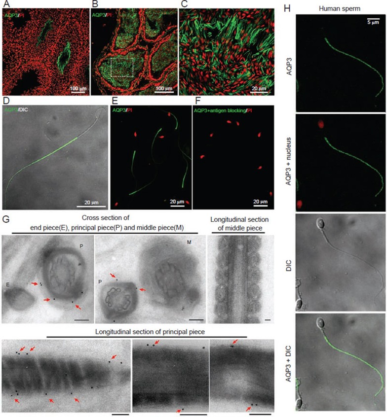
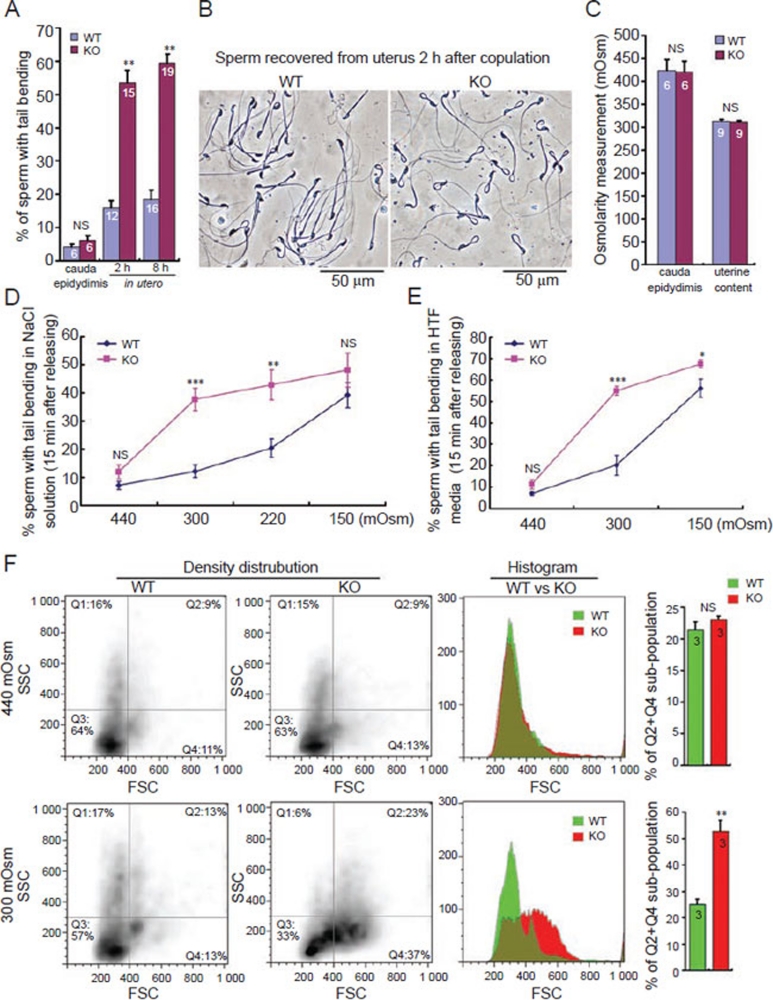
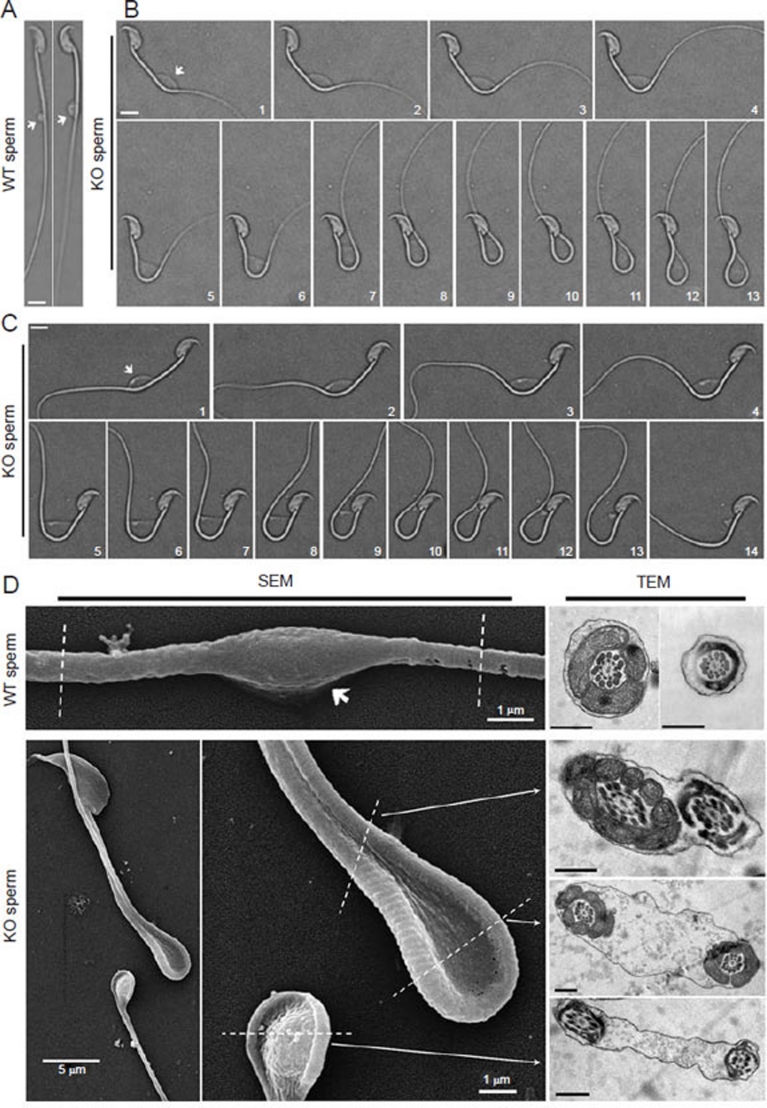
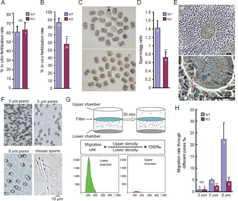
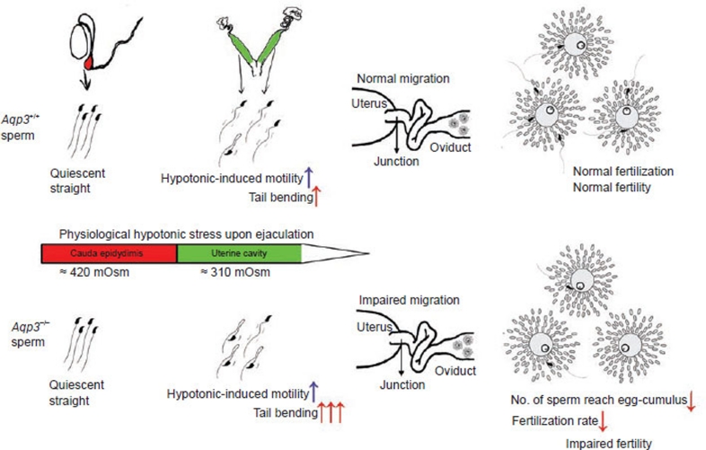
In the journey from the male to female reproductive tract, mammalian sperm experience a natural osmotic decrease (e.g., in mouse, from ~415 mOsm in the cauda epididymis to ~310 mOsm in the uterine cavity). Sperm have evolved to utilize this hypotonic exposure for motility activation, meanwhile efficiently silence the negative impact of hypotonic cell swelling. Previous physiological and pharmacological studies have shown that ion channel-controlled water influx/efflux is actively involved in the process of sperm volume regulation; however, no specific sperm proteins have been found responsible for this rapid osmoadaptation. Here, we report that aquaporin3 (AQP3) is a sperm water channel in mice and humans. Aqp3-deficient sperm show normal motility activation in response to hypotonicity but display increased vulnerability to hypotonic cell swelling, characterized by increased tail bending after entering uterus. The sperm defect is a result of impaired sperm volume regulation and progressive cell swelling in response to physiological hypotonic stress during male-female reproductive tract transition. Time-lapse imaging revealed that the cell volume expansion begins at cytoplasmic droplet, forcing the tail to angulate and form a hairpin-like structure due to mechanical membrane stretch. The tail deformation hampered sperm migration into oviduct, resulting in impaired fertilization and reduced male fertility. These data suggest AQP3 as an essential membrane pathway for sperm regulatory volume decrease (RVD) that balances the "trade-off" between sperm motility and cell swelling upon physiological hypotonicity, thereby optimizing postcopulatory sperm behavior.


Figures

Figure 1
Figure 1
AQP3 is expressed in mouse sperm. (A-D) Immunofluorescence staining of AQP3 in mouse testis (A), cauda epididymis (B, C) and isolated sperm (D) from cauda epididymis. Note the intensive green signal at principal piece of sperm tail. (E, F) AQP3 antibody staining in the absence (E) or presence (F) of competing immunogen. Nucleus was counter stained by propidium iodide (PI). (G) Immunogold-labeled electron microscopic detection shows that gold particles are stained at plasma membrane of the principal piece (indicated by arrows). Scale bars: 0.2 μm. (H) Immunofluorescence detection of AQP3 in human sperm.
Figure 2
Figure 2
Aqp3−/− sperm showed increased in utero tail bending and exaggerated cell swelling upon physiological hypotonic stress. (A) Tail bending of wild-type and Aqp3−/− sperm in cauda epididymis and in postcopulatory uterus (NS: P > 0.05, **P < 0.0001, t-test). (B) Demonstrative pictures of wild-type and Aqp3−/− sperm within uterus at 2 h after copulation. Scale bars: 50 μm. (C) In vivo osmotic environment of wild-type and Aqp3−/− sperm is comparable (NS: P > 0.05, t-test). (D, E) Examination of the sperm tail bending after releasing cauda epididymal sperm into different osmotic environment. NaCl solution (n = 8 for each data presented, NS: P > 0.05, ***P < 0.0001, **P < 0.005. KO vs WT, t-test; D); HTF media (n = 5-8 for each data presented, NS: P > 0.05, ***P < 0.0001, *P < 0.05. KO vs WT, t-test; E). (F) Flow cytometry recorded forward scatter laser (FSC) distribution of the wild-type and Aqp3−/− sperm within different osmolarities (NS: P > 0.05, **P < 0.005, t-test). Numbers within the bars indicate number of mice used for each assay. All error bars represent s.e.m.
Figure 3
Figure 3
Sperm tail bending is caused by mechanical membrane stretch beginning at cytoplasmic droplet. (A-C) Images of wild-type (A) and Aqp3−/− (B, C) sperm from cauda epididymis that were directly released into 300 mOsm NaCl solution. For the Aqp3−/− sperm, time-lapse imaging reveals gradual process of sperm tail bending forced by membrane expansion. Numbers in the picture represent time sequential. Note the swelling state of cytoplasmic droplet (indicated by arrows) in each genotype. Membrane rupture could be clearly observed in 13th photo of (C). Scale bars: 5 μm. (D) Scanning (SEM) and transmission (TEM) electronic microscopy reveal surface and inner appearance of sperm after releasing into 300 mOsm NaCl solution. Note: the bent Aqp3−/− sperm shows expanded intracellular space compared with the wild-type sperm. Scale bars (TEM): 0.5 μm.
Figure 4
Figure 4
Aqp3−/− male shows reduced in vivo fertilization due to impaired sperm migration into oviduct. (A, B) In vitro (A) and in vivo (B) fertilization tests for wild-type and Aqp3−/− sperm (NS: P > 0.05, **P < 0.01, t-test). (C) Representative pictures of embryos flushed from wild-type female oviduct (day 2) after mating with wild-type and Aqp3−/− male. Each picture shows embryos collected from two females. (D) Aqp3−/− sperm shows reduced number in reaching egg-cumulus complex in vivo (**P < 0.01, t-test). (E) Demonstrative figures of egg-cumulus complex (upper) and arrived sperm as indicated by red arrow (lower). Scale bars: 50 μm. (F) Actual size of different pores (3, 5 and 8 μm diameter) in filter membranes (10 μm thick) used for in vitro sperm migration assay. (G) Illustration of in vitro sperm migration assay through filters with different pores and calculation of migration rate. (H) Migration rate for wild-type and Aqp3−/− sperm (released from cauda epididymis) through different filters (NS: P > 0.05, **P < 0.01, KO vs WT, t-test). Numbers within or above the bars indicate number of mice used for each assay. All error bars represent s.e.m.
Figure 5
Figure 5
Schematic figure summarizing the scenario of how AQP3 deficiency impairs male fertility. Upon ejaculation, sperm stored in male reproductive tract enter uterine cavity where they experience a natural osmotic decrease. The physiological hypotonic stress initiates sperm motility, but also poses potential harms to sperm function by inducing sperm cell swelling. Normally, only a small portion of sperm undergoes tail deformation because of the hypotonic stress. However, in Aqp3−/− mice, the Aqp3−/− sperm showed impaired resistance and increased vulnerability to hypotonic-induced cell swelling, resulting in a large portion of sperm showing tail bending after entering uterine environment. The increased tail deformation in the Aqp3−/− sperm led to decreased sperm passage through uterine-oviduct junction and decreased chance for meeting and fertilizing eggs, resulting in impaired male fertility.

References

    1. Morisawa M, Suzuki K. Osmolality and potassium ion: their roles in initiation of sperm motility in teleosts. Science. 1980;210:1145–1147. - PubMed
    1. Cosson J, Groison AL, Suquet M, et al. Marine fish spermatozoa: racing ephemeral swimmers. Reproduction. 2008;136:277–294. - PubMed
    1. Johnson AL, Howards SS. Hyperosmolality in intraluminal fluids from hamster testis and epididymis: a micropuncture study. Science. 1977;195:492–493. - PubMed
    1. Cooper TG, Yeung CH. Acquisition of volume regulatory response of sperm upon maturation in the epididymis and the role of the cytoplasmic droplet. Microsc Res Tech. 2003;61:28–38. - PubMed
    1. Rossato M, Di Virgilio F, Foresta C. Involvement of osmo-sensitive calcium influx in human sperm activation. Mol Hum Reprod. 1996;2:903–909. - PubMed

Publication types

MeSH terms