Review
. 2011 Jan;21(1):131-45.
doi: 10.1038/cr.2010.173. Epub 2010 Dec 7.

Regulation and function of TPL-2, an IκB kinase-regulated MAP kinase kinase kinase

Affiliations
Review

Regulation and function of TPL-2, an IκB kinase-regulated MAP kinase kinase kinase

Thorsten Gantke et al. Cell Res. 2011 Jan.

Erratum in

  • Cell Res. 2011 Apr;21(4):704

Abstract

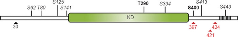
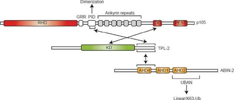
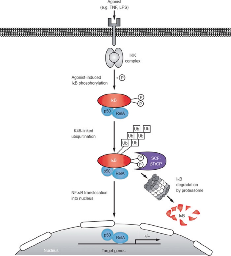
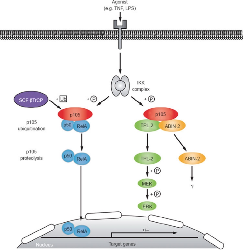
The IκB kinase (IKK) complex plays a well-documented role in innate and adaptive immunity. This function has been widely attributed to its role as the central activator of the NF-κB family of transcription factors. However, another important consequence of IKK activation is the regulation of TPL-2, a MEK kinase that is required for activation of ERK-1/2 MAP kinases in myeloid cells following Toll-like receptor and TNF receptor stimulation. In unstimulated cells, TPL-2 is stoichiometrically complexed with the NF-κB inhibitory protein NF-κB1 p105, which blocks TPL-2 access to its substrate MEK, and the ubiquitin-binding protein ABIN-2 (A20-binding inhibitor of NF-κB 2), both of which are required to maintain TPL-2 protein stability. Following agonist stimulation, the IKK complex phosphorylates p105, triggering its K48-linked ubiquitination and degradation by the proteasome. This releases TPL-2 from p105-mediated inhibition, facilitating activation of MEK, in addition to modulating NF-κB activation by liberating associated Rel subunits for translocation into the nucleus. IKK-induced proteolysis of p105, therefore, can directly regulate both NF-κB and ERK MAP kinase activation via NF-κB1 p105. TPL-2 is critical for production of the proinflammatory cytokine TNF during inflammatory responses. Consequently, there has been considerable interest in the pharmaceutical industry to develop selective TPL-2 inhibitors as drugs for the treatment of TNF-dependent inflammatory diseases, such as rheumatoid arthritis and inflammatory bowel disease. This review summarizes our current understanding of the regulation of TPL-2 signaling function, and also the complex positive and negative roles of TPL-2 in immune and inflammatory responses.


Figures

Figure 1
Figure 1
TPL-2 structure and phosphorylation sites. The Tpl2 gene encodes two proteins, full-length M1-TPL-2 (p58) and M30-TPL-2 (p52). M30-TPL-2 is translated from the same mRNA transcript as M1-TPL-2 by alternative translational initiation at methionine 30 (M30, black arrowhead). The TPL-2 kinase domain (KD) is located in the centre of the protein, flanked by N-terminal and C-terminal regions with largely unknown functions. C-terminal truncation, however, results in a protein (TPL-2ΔC) with increased kinase-specific activity, suggesting that this region may inhibit TPL-2 kinase activity . Furthermore, a proposed degron sequence (435-457, shaded box) is located within the C terminus and confers destabilizing properties to full-length TPL-2 . Consequently, TPL-2ΔC has increased protein stability and is expressed at higher levels. The positions of oncogenic truncations in TPL-2 identified in MoMuLV- and MMTV-induced murine tumors (424), human TPL-2 (COT) in transformed SHOK cells (397) and in a human lung adenocarcinoma (421) are indicated by red arrowheads. Several phosphorylation sites in TPL-2 have been identified by mass spectrometry . Two of these sites, T290 and S400, are known to regulate TPL-2 MEK kinase activity in vivo. T290 phosphorylation may also regulate TPL-2 release from its binding partner p105 (see Figure 4). The physiological significance of the sites shown in italics is not yet known.
Figure 2
Figure 2
TPL-2 interactions with NF-κB1 p105 and ABIN-2. In unstimulated cells, all detectable TPL-2 is complexed with NF-κB1 p105 and ABIN-2. The TPL-2 kinase domain (KD) directly interacts with the death domain (DD) of p105. This regulates TPL-2 MEK kinase activity by blocking access of the substrate to the active site. The TPL-2 C terminus (398-467) interacts with a region (497-539) of p105 within the “processing inhibitory domain” (PID; 474-544) , which also mediates p105 dimerization . These two distinct interactions contribute to a very strong association between TPL-2 and p105, and dissociation of recombinant TPL-2/p105 complex produced in insect cells requires high concentrations of urea (8 M) . The importance of TPL-2 regulation by p105 is highlighted by the dysregulated TPL-2 MEK kinase activity and tumorigenic potential of C-terminal truncated TPL-2, which lacks one of the binding sites for p105 (see text for details). The same region of TPL-2 that mediates binding to the PID of p105 is also the principal interaction site with ABIN-2. The binding site on ABIN-2 (194-250) contains ABIN-homology domain (AHD) 4 (203-220), which is also present in ABIN-1 and ABIN-3 . ABIN-2 interaction is critical to maintain TPL-2 protein stability, and steady-state levels of TPL-2 are dramatically reduced in cells deficient in ABIN-2. RHD: Rel homology domain, GRR: glycine-rich region, PEST: Domain rich in proline (P), glutamate (E), serine (S) and threonine (T), UBAN: ubiquitin-binding in ABIN and NEMO.
Figure 3
Figure 3
Canonical pathway of NF-κB activation. Various stimuli activate the classical NF-κB pathway. Agonist stimulation induces adapter recruitment to the cognate receptor, which then activates the IKK complex by a process that involves linear and K63-linked ubiquitination, as well as phosphorylation (reviewed in Skaug et al. , Vallabhapurapu and Karin ). IKK phosphorylates two N-terminal residues in IκBα, creating a binding site for the SCFβTrCP ubiquitin E3 ligase complex, which attaches K48-linked ubiquitin chains to two adjacent lysine residues, targeting IκBα for proteasomal degradation. Associated NF-κB dimers are consequently liberated to translocate to the nucleus and modulate target gene expression.
Figure 4
Figure 4
Regulation of TPL-2 through IKK-induced p105 proteolysis. TPL-2 is confined to a cytoplasmic complex with the NF-κB1 precursor protein p105, and the ubiquitin-binding protein ABIN-2. NF-κB1 p105 has multiple functions. First, it serves as a precursor molecule for the NF-κB1 p50 subunit, which is generated by limited proteolysis (processing) of p105 by the proteasome, which removes p105's C-terminal half . Second, p105 functions as a classical IκB (see Figure 2), retaining associated NF-κB subunits in the cytoplasm. Cellular stimulation with various agonists, such as TLR ligands, IL-1β, TNF and CD40L, induces the formation of receptor proximal complexes that trigger activation of the MAP3 kinase TAK1 . Activation loop phosphorylation of IKK2 by TAK1, in turn, activates IKK2 to phosphorylate the target residues S927 and S932 in p105, creating a binding site for the SCFβTrCP ubiquitin E3 ligase complex. K48-linked ubiquitination of p105 by SCFβTrCP triggers its complete degradation by the 26S proteasome. IKK-induced proteolysis of p105 releases associated NF-κB dimers, which then translocate to the nucleus and modulate expression of target genes, similar to activation of NF-κB dimers in the classical pathway (see Figure 3). Stimulus-induced p105 proteolysis also couples activation of NF-κB pathways to MAP kinase signaling by releasing TPL-2 from p105 inhibition. After liberation from p105, TPL-2 directly phosphorylates MEK and thereby activates downstream ERK MAP kinase signaling. Based on immunoblotting of total cell lysates, it has been suggested that LPS stimulation results in the selective dissociation of M1-TPL-2 from p105 . However, immunoblotting of p105-depleted lysates indicates that both M1 and M30 TPL-2 are actually released from p105 after LPS stimulation , although the relative contribution of M1 and M30 TPL-2 to MEK phosphorylation in LPS-stimulated macrophages is not known. Activation of TPL-2 MEK kinase activity additionally requires trans-/autophosphorylation of S400 by an unknown kinase and autophosphorylation at T290 in the TPL-2 activation loop. Proteasomal degradation of p105 also releases ABIN-2 from association with p105 and TPL-2. The function and downstream targets of ABIN-2 are not known.

References

    1. Miyoshi J, Higashi T, Mukai H, Ohuchi T, Kakunaga T. Structure and transforming potential of the human cot oncogene encoding a putative protein kinase. Mol Cell Biol. 1991;11:4088–4096. - PMC - PubMed
    1. Patriotis C, Makris A, Bear SE, Tsichlis PN. Tumor progression locus 2 (Tpl-2) encodes a protein kinase involved in the progression of rodent T cell lymphomas and in T cell activation. Proc Natl Acad Sci USA. 1993;90:2251–2255. - PMC - PubMed
    1. Lund AH, Turner G, Trubetskoy A, et al. Genome-wide retroviral insertional tagging of genes involved in cancer in Cdkn2a-deficient mice. Nat Genet. 2002;32:160–165. - PubMed
    1. Mikkers H, Allen J, Knipscheer P, et al. High throughput retroviral tagging to identify components of specific signaling pathways in cancer. Nat Genet. 2002;32:153–159. - PubMed
    1. Erny KM, Peli J, Lambert JF, Muller V, Diggelmann H. Involvement of the TPL-2/COT oncogene in MMTV tumorigenesis. Oncogene. 1996;13:2015–2020. - PubMed

MeSH terms

LinkOut - more resources