Review
. 2011 Mar;121(3):291-312.
doi: 10.1007/s00401-010-0783-x. Epub 2010 Dec 7.

Homeostatic regulation of the endoneurial microenvironment during development, aging and in response to trauma, disease and toxic insult

Affiliations
Review

Homeostatic regulation of the endoneurial microenvironment during development, aging and in response to trauma, disease and toxic insult

Andrew P Mizisin et al. Acta Neuropathol. 2011 Mar.

Abstract

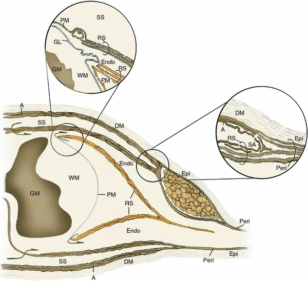
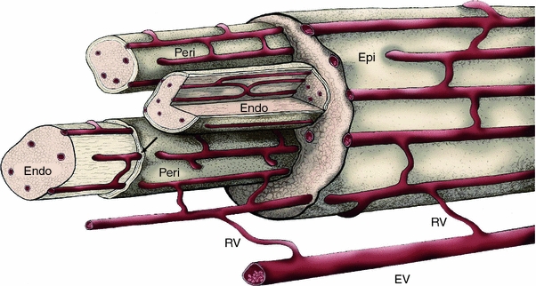
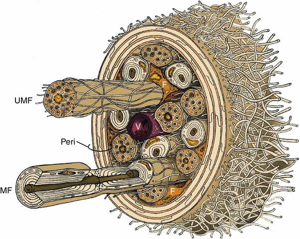
The endoneurial microenvironment, delimited by the endothelium of endoneurial vessels and a multi-layered ensheathing perineurium, is a specialized milieu intérieur within which axons, associated Schwann cells and other resident cells of peripheral nerves function. The endothelium and perineurium restricts as well as regulates exchange of material between the endoneurial microenvironment and the surrounding extracellular space and thus is more appropriately described as a blood-nerve interface (BNI) rather than a blood-nerve barrier (BNB). Input to and output from the endoneurial microenvironment occurs via blood-nerve exchange and convective endoneurial fluid flow driven by a proximo-distal hydrostatic pressure gradient. The independent regulation of the endothelial and perineurial components of the BNI during development, aging and in response to trauma is consistent with homeostatic regulation of the endoneurial microenvironment. Pathophysiological alterations of the endoneurium in experimental allergic neuritis (EAN), and diabetic and lead neuropathy are considered to be perturbations of endoneurial homeostasis. The interactions of Schwann cells, axons, macrophages, and mast cells via cell-cell and cell-matrix signaling regulate the permeability of this interface. A greater knowledge of the dynamic nature of tight junctions and the factors that induce and/or modulate these key elements of the BNI will increase our understanding of peripheral nerve disorders as well as stimulate the development of therapeutic strategies to treat these disorders.


Figures

Fig. 1
Fig. 1
Relationship of the meningeal coverings of the spinal cord to spinal root and peripheral nerve connective tissue ensheathments. The outermost meningeal covering, the dura mater (DM), is continuous with the outermost connective tissue of peripheral nerve, the epineurium (Epi), while the arachnoid layer (A) merges with the outer perineurial lamellae at the subarachnoid angle (SA). The inner layers of the perineurium (Peri) derive from the inner layers of the root sheath (RS). Inset at upper right shows high-power view of the transition of connective tissues at the subarachnoid angle. As the dorsal and ventral spinal roots pass through subarachnoid space (SS), some of the arachnoid layer is reflected onto the root sheath at the subarachnoid angle, becoming the outermost layers of this connective tissue ensheathment. At the root attachment zone of dorsal and ventral roots, the pia mater (PM) of the spinal cord is reflected onto the spinal root and merges with the outer layers of the root sheath, while the glia limitans (GL) continues across the attachment zone to form the interface between the central and peripheral nervous systems. The innermost layers of the root sheath terminate on the spinal root side of the glia limitans. At the root attachment zones, continuity between the subarachnoid space and the endoneurium (Endo) has been demonstrated ultrastructurally (arrows). Inset at upper left illustrates a high-power view of the dorsal root attachment zone, associated spinal cord white matter (WM) and underlying gray matter (GM). Modified from [42, 69]
Fig. 2
Fig. 2
Vasa nervorum. The microcirculation of peripheral nerve derives from regional extrinsic vessels (EV) off of which branch radicular vessels (RV) that supply the intrinsic circulation of the vasa nervorum. The intrinsic circulation consists of longitudinally oriented vessels that course through epineurium (Epi), descend to the perineurium (Peri) and ultimately join with vessels in the endoneurium (Endo) via transperineurial connections (arrow). Extensive anastomotic connections are present at all levels of the intrinsic circulation. Modified from [76]
Fig. 3
Fig. 3
Perineurial ensheathment of peripheral nerve. a A transperineurial vessel in a sural nerve biopsy from diabetic patient has penetrated the outer perineurial lamellae on its way from the epineurium (left) to the endoneurium (right). b Common peroneal nerve biopsy from a diabetic cat showing a portion of fascicle with four perineurial lamellae. Arrow indicates junctional contact between the inner two concentric perineurial cell layers. c Dermal nerve in glabrous skin of rat hind paw ensheathed by a perineurium with only two lamellae. d Perineurium in a common peroneal nerve biopsy from a diabetic cat. Note filaments (black arrows) and dense bodies (white arrow), which are attributes of contractile cells. Bar 6.36 μm in (a), 0.69 μm in (b) 2.00 μm in (c) and 0.27 μm in (d). Electron micrograph in a kindly provided by HC Powell
Fig. 4
Fig. 4
Cellular constituents of the endoneurial microenvironment in peripheral nerve. A small nerve fascicle ensheathed by perineurium (Peri) surrounded by epineurial collagen fibrils is shown with myelinated (MF) and unmyelinated (UMF) fibers, a single endoneurial vessel (V) and scattered fibroblasts (F) embedded in the loose connective tissue of the endoneurium. Not shown are pericytes, resident macrophages and masts cells. Modified from [76]
Fig. 5
Fig. 5
Developmental changes in BNI. Temporal correlation between PSA and 125albumin, BNI index to albumin and endoneurial wet weight/dry weight ratio of rat sciatic nerve during development. Reproduced with permission [189]
Fig. 6
Fig. 6
Endoneurial hydrostatic pressure and perineurial permeability during Wallerian degeneration. BNI PSA to 22Na during the first 4 weeks of Wallerian degeneration is shown. Reproduced with permission [191]
Fig. 7
Fig. 7
Endoneurial fluid pressure and morphometric estimates of endoneurial extracellular space and inflammatory infiltrates in Lewis rats as a function of time after injection with P2-sensitized T cell lines or T cells from rats with adjuvant arthritis (0 days). Note that these parameters all peak at 7 days. Data are presented as mean ± SD and were analyzed with ANOVA after which multiple comparisons were made with the Fisher PLSD test: a P < 0.05 versus 0 days; b P < 0.05 versus 3 days; c P < 0.05 versus 5 days; and d P < 0.05 versus 7 days. Reproduced with permission [131]
Fig. 8
Fig. 8
Tibial nerve endoneurial microvessels in EAN resulting from injection of P2-sensitized T cell lines or T cells from rats with adjuvant arthritis. Vessel from control rat injected with T cells sensitized to adjuvant arthritis (a) or from a rat where EAN was induced by injection of P2-sensitized T cells (b) and (c). Upper inset in a is from control rat and lower inset from a rat with EAN. Arrows indicate interendothelial clefts, which have tight junctions in (a) and are open in (c). Note fragmented endothelial basal lamina in (c) below arrow. Bar 100 μm in insets, 1.43 μm in (a), 2.00 μm in (b) and (c). Reproduced with permission [131]

References

    1. Abbott NJ. Inflammatory mediators and modulation of blood–brain barrier permeability. Cell Mol Neurobiol. 2000;20:131–147. - PMC - PubMed
    1. Abbott NJ. Astrocyte-endothelial interactions and blood–brain barrier permeability. J Anat. 2002;200:629–638. - PMC - PubMed
    1. Aguayo AJ, Charron L, Bray GM. Potential of Schwann cells from unmyelinated nerves to produce myelin: a quantitative ultrastructural and autoradiographic study. J Neurocytol. 1976;5:565–573. - PubMed
    1. Aijaz S, Balda MS, Matter K. Tight junctions: molecular architecture and function. Int Rev Cytol. 2006;248:261–298. - PubMed
    1. Allt G, Lawrenson JG. The blood–nerve barrier: enzymes, transporters and receptors—a comparison with the blood–brain barrier. Brain Res Bull. 2000;52:1–12. - PubMed

Publication types

MeSH terms