Review
. 2011 Mar;68(5):785-801.
doi: 10.1007/s00018-010-0566-5. Epub 2010 Dec 7.

The plasmin-antiplasmin system: structural and functional aspects

Affiliations
Review

The plasmin-antiplasmin system: structural and functional aspects

Johann Schaller et al. Cell Mol Life Sci. 2011 Mar.

Abstract

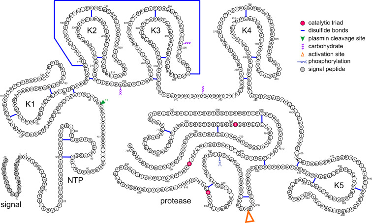
The plasmin-antiplasmin system plays a key role in blood coagulation and fibrinolysis. Plasmin and α(2)-antiplasmin are primarily responsible for a controlled and regulated dissolution of the fibrin polymers into soluble fragments. However, besides plasmin(ogen) and α(2)-antiplasmin the system contains a series of specific activators and inhibitors. The main physiological activators of plasminogen are tissue-type plasminogen activator, which is mainly involved in the dissolution of the fibrin polymers by plasmin, and urokinase-type plasminogen activator, which is primarily responsible for the generation of plasmin activity in the intercellular space. Both activators are multidomain serine proteases. Besides the main physiological inhibitor α(2)-antiplasmin, the plasmin-antiplasmin system is also regulated by the general protease inhibitor α(2)-macroglobulin, a member of the protease inhibitor I39 family. The activity of the plasminogen activators is primarily regulated by the plasminogen activator inhibitors 1 and 2, members of the serine protease inhibitor superfamily.


Figures

Fig. 1
Fig. 1
Schematic representation of the main components of the fibrinolytic system (adapted from reference [2]). sc-tPA, tc-tPA single-chain, two-chain tPA; sc-uPA, tc-uPA single-chain, two-chain uPA; PAIs plasminogen activator inhibitors
Fig. 2
Fig. 2
Schematic representation of the primary structure of human Pgn (from reference [2]). The catalytic triad (His603, Asp646, and Ser741), the activation site (Arg561–Val562), the Plm cleavage site (Lys77–Lys78), the phosphorylation site (Ser578), the CHO attachment sites (Asn289, Ser249, and Thr346), and the 24 disulfide bridges as well as the signal peptide are indicated. NTP N-terminal peptide; K1–K5 kringles 1–5
Fig. 3
Fig. 3
Top and bottom 3-D structural model of human Pgn based on overlapping 3-D structures of Pgn fragments (form reference [44]). Each kringle is shown in grey, the central Trp residue in each LBS is shown in magenta, the protease domain is shown in green, and the active site residues are shown in red
Fig. 4
Fig. 4
3-D structure of human Pgn kringles 1 + 2 + 3 (K1, K2, K3) termed angiostatin determined by X-ray diffraction (1KI0, [47]). Each kringle contains three disulfide bridges (yellow) arranged in the pattern Cys1–Cys6, Cys2–Cys4, Cys3–Cys5 exemplified in kringle 2 (K2). Kringle 1 (K1) is shown as a surface representation with negatively and positively charged residues shown in red and blue, respectively, and all other residues are shown in grey. The central Trp residue of the LBS is shown in magenta (arrow)
Fig. 5
Fig. 5
3-D structure of the catalytic domain of human Pgn determined by X-ray diffraction (1DDJ, [25]). The active site residues located at the interface of the two structurally similar subdomains are shown in magenta
Fig. 6
Fig. 6
Cleavage of human fibrinogen by Plm (from reference [48]). Schematic representation of fibrinogen, the fibrin polymer, the pattern of cleavage by Plm, and the main fragments generated. The main Plm cleavage sites are indicated by arrows. Cleavage of fibrin by Plm leads to the main fragments X (260 kDa), Y (160 kDa), D (100 kDa), and E (60 kDa)
Fig. 7
Fig. 7
a 3-D structure of the double module of human tPA comprising the fibronectin type I (FN1) domain and the epidermal growth factor-like (EGF-like) domain determined by NMR spectroscopy (1TPG, [74]). The disulfide bridges shown in yellow exhibit the following patterns: in FN1 (red) Cys1–Cys3, Cys2–Cys4, and in EGF-like (blue) Cys1–Cys3, Cys2–Cys4, Cys5–Cys6. b 3-D structure of the catalytic domain of human tPA in complex with the LMW inhibitor dansyl-EGR-chloromethyl ketone determined by X-ray diffraction (1BDA, [78]). The inhibitor shown in red is covalently bound to His322 and Ser478 in the active site cleft (magenta)
Fig. 8
Fig. 8
a 3-D structure of the EGF-like (blue) and kringle (green) double domain of human uPA determined by NMR spectroscopy (1URK, [92]). The disulfide bridges are shown in yellow and are arranged as in Fig. 7a (EGF-like) and Fig. 4 (kringle). b 3-D structure of human LMW uPA determined by X-ray diffraction (1GJA, [96]). The minichain (red) is linked to the catalytic domain by a single interchain disulfide bridge (yellow)
Fig. 9
Fig. 9
Various conformational states of serpins [100]. a Native state of human α1-antitrypsin (1QLP). b Latent, inserted state of human antithrombin III (2ANT). c Cleaved state of human α1-antitrypsin (7API). d Noncovalent, Michaelis-like complex of alaserpin (from Manduca sexta) in complex with rat trypsin (1I99). e Cleaved serpin with inactivated serine protease of human α1-antitrypsin with bovine trypsin (1EZX)
Fig. 10
Fig. 10
3-D structure of human PAI-1 in complex with two inhibitory RCL pentapeptides (Ac-TVASS-NH2) determined by X-ray diffraction (1A7C, [144]). The two pentapeptides (blue) bind between β-strands 3 and 5 in β-sheet A (red). The cleaved C-terminal region is shown in magenta and the cleaved ends of the reactive peptide bond are located on either side of the molecule (red circles)
Fig. 11
Fig. 11
3-D structure of a deletion mutant of human PAI-2 determined by X-ray diffraction (1BY7, [171]). The reconstructed RCL is shown as a dashed blue line and the shutter region located beneath β-sheet A (red) is shown as a blue-shaded oval
Fig. 12
Fig. 12
3-D structure of uncleaved, native human neuroserpin determined by X-ray diffraction (3F5N, 3F02, [180]). Neuroserpin contains the typical structural elements of uncleaved, native serpins: three β-sheets (red, green, yellow), nine α-helices (grey), and the exposed RCL (blue)

References

    1. Schaller J, Gerber S, Kämpfer U, Lejon S, Trachsel C. Human blood plasma proteins: structure and function. Chichester: Wiley; 2008.
    1. Gerber SS (2009) The human α2-plasmin inhibitor: functional characterization of the unique plasmin(ogen)-binding region. Inaugural dissertation, University of Bern, Switzerland
    1. Waisman DM. Plasminogen: structure, activation, and regulation. New York: Kluwer Academic/Plenum Publishers; 2003.
    1. Syrovets T, Simmet T. Novel aspects and new roles of the serine protease plasmin. Cell Mol Life Sci. 2004;61:873–885. doi: 10.1007/s00018-003-3348-5. - DOI - PMC - PubMed
    1. Myöhänen H, Vaheri A. Regulation and interaction in the activation of cell-associated plasminogen. Cell Mol Life Sci. 2004;61:2840–2858. doi: 10.1007/s00018-004-4230-9. - DOI - PMC - PubMed

MeSH terms