. 2010 Dec 7:5:55.
doi: 10.1186/1750-1326-5-55.

α-synuclein induced synapse damage is enhanced by amyloid-β1-42

Affiliations

α-synuclein induced synapse damage is enhanced by amyloid-β1-42

Clive Bate et al. Mol Neurodegener. .

Abstract

Background: The pathogenesis of Parkinson's disease (PD) and dementia with Lewy bodies (DLB) is associated with the accumulation of aggregated forms of the α-synuclein (αSN) protein. An early event in the neuropathology of PD and DLB is the loss of synapses and a corresponding reduction in the level of synaptic proteins. However, the molecular mechanisms involved in synapse damage in these diseases are poorly understood. In this study the process of synapse damage was investigated by measuring the amount of synaptophysin, a pre-synaptic membrane protein essential for neurotransmission, in cultured neurons incubated with αSN, or with amyloid-β (Aβ) peptides that are thought to trigger synapse degeneration in Alzheimer's disease.

Results: We report that the addition of recombinant human αSN reduced the amount of synaptophysin in cultured cortical and hippocampal neurons indicative of synapse damage. αSN also reduced synaptic vesicle recycling, as measured by the uptake of the fluorescent dye FM1-43. These effects of αSN on synapses were modified by interactions with other proteins. Thus, the addition of βSN reduced the effects of αSN on synapses. In contrast, the addition of amyloid-β (Aβ)1-42 exacerbated the effects of αSN on synaptic vesicle recycling and synapse damage. Similarly, the addition of αSN increased synapse damage induced by Aβ1-42. However, this effect of αSN was selective as it did not affect synapse damage induced by the prion-derived peptide PrP82-146.

Conclusions: These results are consistent with the hypothesis that oligomers of αSN trigger synapse damage in the brains of Parkinson's disease patients. Moreover, they suggest that the effect of αSN on synapses may be influenced by interactions with other peptides produced within the brain.


Figures
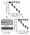
Figure 1
Figure 1
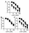
αSN triggered the loss of synaptophysin from cultured neurons. (A) The synaptophysin content of cortical neurons incubated for 24 hours with αSN (●) or βSN (○) as shown. Values shown are the mean amount of synaptophysin (units) ± SD, n = 15. (B) Immunoblots showing the amount of synaptophysin and β-actin in extracts from cortical neurons that had been incubated for 24 hours with αSN as shown. (C) The synaptophysin content of hippocampal neurons incubated for 24 hours with αSN (●) or βSN (○) as shown. Values shown are the mean amount of synaptophysin (units) ± SD, n = 12.
Figure 2
Figure 2
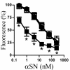
αSN reduced synaptic vesicle recycling. The amount of the fluorescent dye (FM1-43) taken up by synaptic vesicles in cortical neurons that had been incubated for 24 hours with different concentrations of αSN (●) or βSN (○) as shown and stimulated with ACh for 10 minutes. Values shown are the mean % fluorescence (where 100% = fluorescence in control cortical neurons) ± SD, n = 15.
Figure 3
Figure 3
βSN reduced the αSN-induced loss of synaptophysin. The synaptophysin content of cortical neurons incubated with varying concentrations of αSN (●), αSN premixed with βSN (1:10)(□) or αSN premixed with human serum albumin (1:10)(■) for 24 hours. Values shown are the mean amount of synaptophysin (units) ± SD, n = 12. (*) = amount of synaptophysin significantly higher than in neurons incubated with the same concentration of αSN alone (P < 0.01).
Figure 4
Figure 4
1-42 enhanced the αSN-induced loss of synaptophysin. (A) The synaptophysin content of cortical neurons incubated for 24 hours with varying concentrations of αSN (●), αSN premixed with Aβ1-42 (50:1) (■) or αSN premixed with Aβ42-1 (50:1) (□). Values shown are the mean amount of synaptophysin (units) ± SD, n = 18. (*) = amount of synaptophysin significantly lower than in neurons incubated with the same concentration of αSN alone (P < 0.01). (B) The synaptophysin content of cortical neurons pre-treated for 1 hour with control medium (●) or 1 nM Aβ1-42 (○) and incubated for 24 hours with αSN as shown. Values shown are the mean amount of synaptophysin (units) ± SD, n = 12. (C) The synaptophysin content of cortical neurons incubated for 24 hours with αSN (●), αSN premixed with 7PA2-CM (■) or αSN premixed with CHO-CM (□). Values shown are the mean amount of synaptophysin (units) ± SD, n = 12. (*) = amount of synaptophysin significantly lower than in neurons incubated with the same concentration of αSN alone (P < 0.01).
Figure 5
Figure 5
1-42 enhanced the αSN-induced inhibition of synaptic vesicle recycling. The amount of the fluorescent dye (FM1-43) taken up into synaptic vesicles in cortical neurons that had been incubated for 24 hours with different concentrations of αSN (●), or with αSN that had been premixed with either 7PA2-CM (■) or with CHO-CM (□) as shown and stimulated with ACh for 10 minutes. Values shown are the mean % fluorescence (where 100% = fluorescence in vehicle treated cortical neurons) ± SD, n = 18. (*) = Fluorescence significantly lower than in neurons incubated with the same concentration of αSN alone (P < 0.01).
Figure 6
Figure 6
αSN enhanced the Aβ1-42-induced loss of synaptophysin. (A) The synaptophysin content of cortical neurons incubated for 24 hours with Aβ1-42 (●) Aβ1-42 premixed with αSN (1:2) (■) or Aβ1-42 premixed with βSN (1:2) (□). Values shown are the mean amount of synaptophysin (units) ± SD, n = 15. (*) = amount of synaptophysin significantly lower than in neurons incubated with the same concentration of Aβ1-42 alone (P < 0.01). (B) The synaptophysin content of cortical neurons incubated for 24 hours with PrP82-146 (●) or PrP82-146 that had been premixed with αSN (1:5) (○). Values shown are the mean amount of synaptophysin (units) ± SD, n = 15.
Figure 7
Figure 7
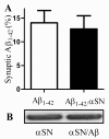
αSN did not affect the accumulation of Aβ1-42 in synapses. (A) The amount of biotinylated-Aβ1-42 found in synaptosomes derived from cortical neurons incubated for 1 hour with 100 nM Aβ1-42 (□) or 100 nM Aβ1-42 that had been pre-mixed with 500 nM αSN (■). Values shown are the mean amount of Aβ1-42 expressed as a % of the amount added ± SD, n = 12. (B) Immunoblot showing the amount of αSN in synaptosomes collected from cortical neurons incubated with 200 nM αSN alone or with 200 nM αSN pre-mixed with Aβ1-42 (50:1).

References

    1. Poewe W. Non-motor symptoms in Parkinson's disease. Eur J Neurol. 2008;15(Suppl 1):14–20. doi: 10.1111/j.1468-1331.2008.02056.x. - DOI - PubMed
    1. Aarsland D, Andersen K, Larsen JP, Lolk A, Kragh-Sorensen P. Prevalence and characteristics of dementia in Parkinson disease: an 8-year prospective study. Arch Neurol. 2003;60(3):387–392. doi: 10.1001/archneur.60.3.387. - DOI - PubMed
    1. McKeith IG, Dickson DW, Lowe J, Emre M, O'Brien JT, Feldman H, Cummings J, Duda JE, Lippa C, Perry EK. et al.Diagnosis and management of dementia with Lewy bodies: third report of the DLB Consortium. Neurology. 2005;65(12):1863–1872. doi: 10.1212/01.wnl.0000187889.17253.b1. - DOI - PubMed
    1. Bonini NM, Giasson BI. Snaring the function of alpha-synuclein. Cell. 2005;123(3):359–361. doi: 10.1016/j.cell.2005.10.017. - DOI - PubMed
    1. Cabin DE, Shimazu K, Murphy D, Cole NB, Gottschalk W, McIlwain KL, Orrison B, Chen A, Ellis CE, Paylor R. et al.Synaptic vesicle depletion correlates with attenuated synaptic responses to prolonged repetitive stimulation in mice lacking alpha-synuclein. J Neurosci. 2002;22(20):8797–8807. - PMC - PubMed

LinkOut - more resources