. 2011 Feb 25;286(8):6165-74.
doi: 10.1074/jbc.M110.142992. Epub 2010 Dec 6.

Determinants of the specificity of rotavirus interactions with the alpha2beta1 integrin

Affiliations

Determinants of the specificity of rotavirus interactions with the alpha2beta1 integrin

Fiona E Fleming et al. J Biol Chem. .

Abstract

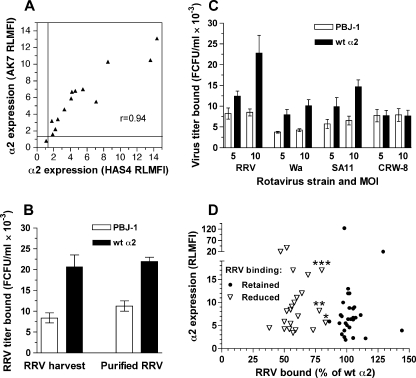
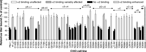
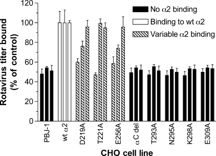
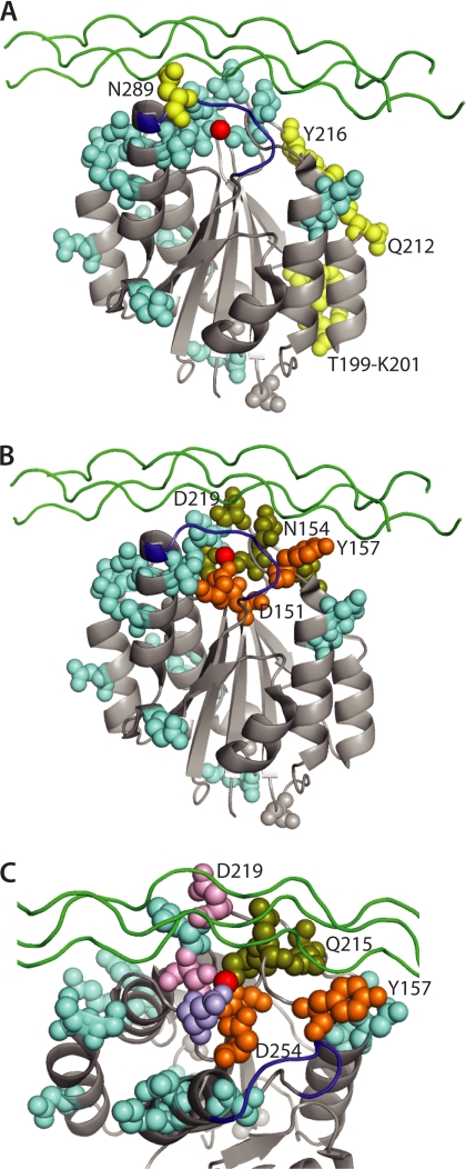
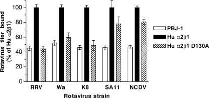
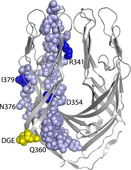
The human α2β1 integrin binds collagen and acts as a cellular receptor for rotaviruses and human echovirus 1. These ligands require the inserted (I) domain within the α2 subunit of α2β1 for binding. Previous studies have identified the binding sites for collagen and echovirus 1 in the α2 I domain. We used CHO cells expressing mutated α2β1 to identify amino acids involved in binding to human and animal rotaviruses. Residues where mutation affected rotavirus binding were located in several exposed loops and adjacent regions of the α2 I domain. Binding by all rotaviruses was eliminated by mutations in the activation-responsive αC-α6 and αF helices. This is a novel feature that distinguishes rotavirus from other α2β1 ligands. Mutation of residues that co-ordinate the metal ion (Ser-153, Thr-221, and Glu-256 in α2 and Asp-130 in β1) and nearby amino acids (Ser-154, Gln-215, and Asp-219) also inhibited rotavirus binding. The importance of most of these residues was greatest for binding by human rotaviruses. These mutations inhibit collagen binding to α2β1 (apart from Glu-256) but do not affect echovirus binding. Overall, residues where mutation affected both rotavirus and collagen recognition are located at one side of the metal ion-dependent adhesion site, whereas those important for collagen alone cluster nearby. Mutations eliminating rotavirus and echovirus binding are distinct, consistent with the respective preference of these viruses for activated or inactive α2β1. In contrast, rotavirus and collagen utilize activated α2β1 and show an overlap in α2β1 residues important for binding.


Figures

FIGURE 1.
FIGURE 1.
Determining the parameters of rotavirus binding to native and mutant α2 on CHO cells. A, levels of α2 expression determined by flow cytometry using AK7 and HAS4 antibodies, expressed as RLMFI, were compared using cells expressing empty vector (PBJ-1), wt human α2 (Hu α2), wt human α2β1, and mutated human α2 (randomly selected; Y157A, Q212A, R213A, G240A, D259A, K264A, D273A, L291A, D292A, K298A, E309A). Lines indicate the positive-negative RLMFI cut-off of 1.2. PBJ-1 cells exhibited the single negative RLMFI value. B, RRV purification did not alter virus titers bound to PBJ-1 and Hu α2 CHO cells. RRV cell culture harvest (RRV harvest) and purified RRV were bound to cells at a multiplicity of infection of 10. C, dose-dependence of rotavirus binding to α2β1 on CHO cells. Binding of representative rotavirus strains RRV, Wa, SA11, and CRW-8 was evaluated at a multiplicity of infection (MOI) of 5 and 10. Data obtained with rotaviruses K8 and NCDV were similar to Wa binding levels. D, comparison of α2 RLMFI values and RRV binding levels for cells expressing mutated α2 that retained RRV binding (n = 29) or showed reduced RRV binding (n = 22, comprising 21 point mutants and the αC deletion mutant). RLMFI values for α2 were determined using AK7 except for CHO α2 R288A, which was recognized by HAS4 only. Data marked *, **, and *** represent CHO α2 E256A, N295A, and K163A, respectively. RRV and Wa bound these mutants at intermediate and low levels, respectively (Fig. 2).
FIGURE 2.
FIGURE 2.
Effect of mutations in α2I on α2β1 binding by rotavirus strains RRV and Wa. CHO cells stably expressing empty vector (PBJ-1), wild-type human α2 (wt α2), or mutant human α2 were used to determine binding by monkey rotavirus RRV (first bar in each pair, or single bar) and human rotavirus Wa (second bar of each pair). The mutations consisted of single amino acid changes as indicated, with the exception of αC del that contained a deletion of the αC loop (amino acids 283–290). Data are presented as a percentage of the virus titer bound to wt α2 cells. These cells bound twice the level of virus bound by cells containing the empty vector. As cellular α2 expression was similar between mutant cell lines bound by rotaviruses and those that did not bind virus (Fig. 1), these data were not normalized for α2 expression levels. Secondary structural elements of α2I (α-helices and β-sheets as defined in Ref. 34) are indicated above the bars.
FIGURE 3.
FIGURE 3.
Analysis of binding by rotaviruses K8 (human), SA11 (monkey), and NCDV (bovine) to α2I domain mutants that variably affected RRV and Wa binding, and α2I containing an αC loop deletion (amino acids 283–290) or single α6 loop mutations. Rotaviruses were bound to CHO cell lines selected from those described in the legend to Fig. 2. Titers bound by K8, SA11, and NCDV are represented by the first, second, and third bars in each group, respectively. Data are presented as a percentage of the virus titer bound to CHO parental cells expressing wild-type human α2 (wt α2).
FIGURE 4.
FIGURE 4.
Effect of human-to-mouse swapping and point mutations in α2I on rotavirus binding and infection through cell surface-expressed α2β1. Relative levels of binding by rotaviruses RRV and Wa (A) and K8, SA11, and NCDV (B) to CHO cells expressing empty vector (PBJ-1), human α2 (Hu α2), mouse α2I within human α2 (Mur α2I), or human α2 with amino acids 212–216 swapped to the mouse sequence (Hu-mur α2I) are illustrated. C, infectious rotavirus titers produced at 16 h after infection of PBJ-1, Hu α2, and Hu-mur α2I by RRV, Wa, NCDV, or integrin-independent porcine rotavirus CRW-8 at a multiplicity of infection of 1. D, infectious RRV titers produced at 16 h after infection of PBJ-1, Hu α2, Y157A, N289A, T293A, and D243A.
FIGURE 5.
FIGURE 5.
Structural comparison of the α2I locations of amino acids affecting rotavirus, EV1, and type I collagen binding. The sites of mutations inhibiting rotavirus or EV1 binding (A), and rotavirus and/or collagen binding (B, C) are compared individually, with the atoms of residues where mutation affects binding shown as colored-filled spheres. The view in A and B is identical, looking from one side with the MIDAS at the top. In C, the view is from the same side from the top and is restricted to the MIDAS and surrounds. Mutations affecting the binding of rotaviruses (light blue), echovirus (yellow), collagen but not viruses (orange), and both rotaviruses and collagen (olive green) are indicated. In C, amino acids with mutations that affected collagen binding and variably affected rotavirus binding (Asp-219, Thr-221) are shown in pink, and Glu-256 where mutation variably affected rotavirus binding only is shown in purple. In all panels the MIDAS divalent cation is shown in red, the position of a bound model triple-helical collagen peptide is given in green, and the αC loop is shown in dark blue. This is adapted from the x-ray structure of activated α2I bound to triple-helical collagen peptide, Protein DataBase Code 1DZI (21).
FIGURE 6.
FIGURE 6.
Effect of D130A mutation in the β1 subunit of human α2β1 on rotavirus binding to cell surface-expressed α2β1. Relative infectious titers of RRV, Wa, K8, SA11, and NCDV bound to CHO cells transfected with human α2β1 (Hu α2β1), or human α2β1 with the point mutation D130A in the β1 subunit (Hu α2β1 D130A) are given. The titer of RRV and SA11 bound to Hu α2β1 cells is increased by 22 and 27%, respectively, over that bound to wt α2 cells (17).
FIGURE 7.
FIGURE 7.
Location of DGE sequence (yellow) and residues 335 to 380 (pale blue) in the truncated RRV dimeric VP5* structure considered to represent the protease-activated form. Residues differing between the RRV/Wa/K8 and SA11/NCDV rotavirus clusters are indicated in dark blue. The VP5* dimer is oriented with the hydrophobic loops required for cell membrane disruption at the top of the image. This is adapted from the x-ray structure of dimeric RRV VP5*, Protein DataBase Code 2B4H (63).

References

    1. Hynes R. O. (2002) Cell 110, 673–687 - PubMed
    1. Stewart P. L., Nemerow G. R. (2007) Trends Microbiol. 15, 500–507 - PubMed
    1. Triantafilou K., Takada Y., Triantafilou M. (2001) Crit. Rev. Immunol. 21, 311–322 - PubMed
    1. Greenberg H. B., Estes M. K. (2009) Gastroenterology 136, 1939–1951 - PMC - PubMed
    1. Ciarlet M., Crawford S. E., Cheng E., Blutt S. E., Rice D. A., Bergelson J. M., Estes M. K. (2002) J. Virol. 76, 1109–1123 - PMC - PubMed

Publication types

Substances