. 2011 Mar 1;20(5):867-79.
doi: 10.1093/hmg/ddq526. Epub 2010 Dec 6.

PINK1 cleavage at position A103 by the mitochondrial protease PARL

Affiliations

PINK1 cleavage at position A103 by the mitochondrial protease PARL

Emma Deas et al. Hum Mol Genet. .

Abstract

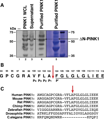
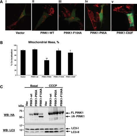
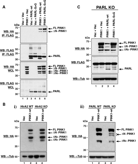
Mutations in PTEN-induced kinase 1 (PINK1) cause early onset autosomal recessive Parkinson's disease (PD). PINK1 is a 63 kDa protein kinase, which exerts a neuroprotective function and is known to localize to mitochondria. Upon entry into the organelle, PINK1 is cleaved to produce a ∼53 kDa protein (ΔN-PINK1). In this paper, we show that PINK1 is cleaved between amino acids Ala-103 and Phe-104 to generate ΔN-PINK1. We demonstrate that a reduced ability to cleave PINK1, and the consequent accumulation of full-length protein, results in mitochondrial abnormalities reminiscent of those observed in PINK1 knockout cells, including disruption of the mitochondrial network and a reduction in mitochondrial mass. Notably, we assessed three N-terminal PD-associated PINK1 mutations located close to the cleavage site and, while these do not prevent PINK1 cleavage, they alter the ratio of full-length to ΔN-PINK1 protein in cells, resulting in an altered mitochondrial phenotype. Finally, we show that PINK1 interacts with the mitochondrial protease presenilin-associated rhomboid-like protein (PARL) and that loss of PARL results in aberrant PINK1 cleavage in mammalian cells. These combined results suggest that PINK1 cleavage is important for basal mitochondrial health and that PARL cleaves PINK1 to produce the ΔN-PINK1 fragment.


Figures

Figure 1.
Figure 1.
Isolation of ΔN-PINK1 and identification of the cleavage site. (A) WB analysis of vector control-transfected HEK293T cells (lane 1), PINK1-3xHA expression in whole-cell lysate samples before purification with anti-HA beads (lane 2), the presence of residual unbound PINK1-3xHA in supernatant post-purification (lane 3) and enrichment of the ΔN-PINK1 protein in the purified sample (lane 4). WBs were performed using an anti-HA antibody. Lane 5 displays the presence of the ΔN-PINK1 protein assessed by Coomassie staining. This membrane was then sent for N-terminal Edman degradation sequencing analysis. (B) N-terminal Edman sequencing result. The 10 amino acids underlined display the amino acid read obtained during the analysis. The sequence of PINK1 spanning amino acids 94–112 is shown with the cleavage site highlighted by a red arrow. The P4–P1 and P′ sites of the PINK1 cleavage site are also indicated. (C) Sequence alignment of the PINK1 protein surrounding the cleavage site showing conservation between mammalian species. Alignment was performed in Clustal X.
Figure 2.
Figure 2.
Efficient PINK1 cleavage is required for mitochondrial health. (A) Cleavage status of the PINK1 cleavage mutants compared with PINK1-wt. Vector control, PINK1-wt-3xHA, PINK1-P95A-3xHA, PINK1-F104A and PINK1-F104D constructs were expressed in SH-SY5Ys. Lysates were then assessed by WB analysis using an anti-HA antibody. (B) PINK1 cleavage mutants still localize to mitochondria. Vector control, PINK1-wt-3xHA, PINK1-P95A-3xHA and PINK1-F104A constructs were expressed in SH-SY5Y cells. Cell lysates were fractionated to isolate cytoplasmic and crude mitochondria. The presence of the PINK1 cleavage mutants in cytosolic and mitochondrial fractions was assessed by WB analysis using an anti-HA antibody. The membrane was subsequently re-probed with anti-ApoTrack cocktail which demonstrates the purity of the fractions and equal loading. (C) Cleavage of the N-terminal PINK1 PD mutant proteins. SH-SY5Y lysates expressing PINK1-C92F, PINK1-Q115L and PINK1-R147H constructs were analysed by WB with an anti-HA antibody. (D) (i) The cleavage status of PINK1 has important effects on basal mitochondrial membrane potential. Failure to cleave the PINK1 protein efficiently, shown through expression of the PINK1-P95A mutant, induces a significant reduction in mitochondrial membrane potential and makes cells more susceptible to stress-mediated death. Vector-transfected control cells were taken to be 100%. *P< 0.05 and **P< 0.005. (ii) A reduced ability to cleave PINK1 results in a significant increase in basal cytoplasmic ROS production. Basal ROS production in vector-transfected control cells was taken as 100%. **P< 0.005. (iii) Impaired PINK1 processing additionally results in a significant increase in basal mitochondrial ROS production. Basal ROS production in vector control-transfected cells was taken as 100%. **P< 0.005. (E) Assessment of ROS production in the PINK1-P95A and PINK1-F104A cleavage mutants compared with PINK1-wt. Basal ROS production increases over time in cells expressing the PINK1-P95A mutant without any stimulus. Stimulation of ROS production using rotenone to block complex 1 function has no effect on ROS production in PINK1-P95A cells but dramatically increases ROS production in both PINK1-wt and PINK1-F104A cells.
Figure 3.
Figure 3.
PINK1 cleavage status affects the mitochondrial network and mass. (A) Failure to cleave PINK1 results in an abnormal mitochondrial network and altered location. Images display SH-SY5Y cells expressing the indicated PINK1 pIRES constructs (green cells) and neighbouring untransfected control cells where only mitochondria are visible. Image (i) shows the normal mitochondrial network observed in cells expressing vector control; (ii) PINK1-wt; (iii) PINK1-F104A; (iv) the loss of mitochondrial network and presence of aggregated mitochondria in cells expressing PINK1-P95A; (v) the intermediate phenotype between PINK1-wt and PINK1-P95A observed in cells expressing the PD PINK1-C92F mutant protein. Mitochondria were visualized using TMRM dye. 3D movies are provided in Supplementary Material, Figures S1–S5. (B) Mitochondrial mass appears reduced in cells predominantly expressing FL-PINK1. Cells expressing PINK1-P95A or PINK1-C92F PD mutant protein show significantly reduced mitochondrial mass compared with vector-, PINK1-wt- and PINK1-F104A-expressing cells. Vector control-transfected cells were taken as 100%. (C) Loss of mitochondrial mass through expression of PINK1-P95A does not correlate with autophagy induction. Cells stably expressing either vector control, PINK1-wt, PINK1-P95A or PINK1-F104A were exposed to DMSO control or CCCP for 12 h prior to lysis. Lysates were analysed by WB with an anti-HA antibody (PINK1) and LC3 antibody to detect autophagy activation by LC3I-II cleavage. Expression of PINK1-P95A did not induce an increase in LC3I-II cleavage at basal levels compared with vector control, PINK1-wt or PINK1-F104A cells. CCCP treatment of all cell lines induced LC3I-II cleavage and suppressed PINK1 cleavage in all cell lines.
Figure 4.
Figure 4.
PINK1 interacts with and is cleaved by PARL. (A) Co-immunoprecipitation of PINK1 with both PARL-wt and the catalytically inactive PARL-S277G mutant. Whole-cell lysates (WCL) from HEK293T cells transiently expressing PINK1-wt-3xHA, PARL-wt-FLAG and PARL-S277G-FLAG were immunoprecipitated (IP) with anti-FLAG® M2-agarose affinity gel prior to analysis by WB with the indicated antibodies. (B) (i) PINK1-wt cleavage status in HtrA2 KO and WT MEFs. HtrA2 KO and WT MEFs were transfected with PINK1-wt-3xHA. Lysates were then assessed by WB analysis using an anti-HA antibody. (ii) PINK1-wt cleavage status in PARL KO and WT MEFs. PARL KO and WT MEFs were transfected with PINK1-wt-3xHA. Lysates were then assessed by WB analysis using an anti-HA antibody. (C) PINK1 cleavage can be restored in PARL KO cells overexpressing PARL-wt but not the catalytically inactive PARL-S277G mutant. PARL KO MEFs were transfected with PINK1-wt-3xHA, PARL-wt-FLAG and PARL-S277G-FLAG. Lysates were then analysed by WB with the indicated antibodies.
Figure 5.
Figure 5.
The PINK1-P95A mutation does not abrogate the PINK1–PARL interaction. (A) Alterations in PINK1 cleavage induced by P95A and F104A mutations are suppressed by the loss of PARL. Vector control, PINK1-wt, PINK1-P95A and PINK1-F104A were expressed in PARL WT and KO MEFs. Lysates were assessed by WB analysis using an anti-HA antibody. (B) PINK1-P95A still interacts with PARL-wt or PARL-S277G. Lysates from HEK293T cells expressing PINK1-wt-3xHA, PINK1-P95A-3xHA, PARL-wt-FLAG and PARL-S277G-FLAG were immunoprecipitated (IP) with anti-FLAG® M2-agarose affinity gel prior to analysis by WB with the indicated antibodies. (C) Structural predictions of the PINK1-wt TM domain and alterations made by the P95A mutation. (D) Assessment of ROS production in cells expressing vector control, PINK1-wt or PINK1-P95A in a PARL RNAi or scrambled RNAi control background.

References

    1. Ibanez P., Lesage S., Lohmann E., Thobois S., De Michele G., Borg M., Agid Y., Durr A., Brice A. Mutational analysis of the PINK1 gene in early-onset parkinsonism in Europe and North Africa. Brain. 2006;129:686–694. - PubMed
    1. Valente E.M., Abou-Sleiman P.M., Caputo V., Muqit M.M., Harvey K., Gispert S., Ali Z., Del Turco D., Bentivoglio A.R., Healy D.G., et al. Hereditary early-onset Parkinson's disease caused by mutations in PINK1. Science. 2004;304:1158–1160. - PubMed
    1. Sim C.H., Lio D.S., Mok S.S., Masters C.L., Hill A.F., Culvenor J.G., Cheng H.C. C-terminal truncation and Parkinson's disease-associated mutations down-regulate the protein serine/threonine kinase activity of PTEN-induced kinase-1. Hum. Mol. Genet. 2006;15:3251–3262. - PubMed
    1. Haque M.E., Thomas K.J., D'Souza C., Callaghan S., Kitada T., Slack R.S., Fraser P., Cookson M.R., Tandon A., Park D.S. Cytoplasmic Pink1 activity protects neurons from dopaminergic neurotoxin MPTP. Proc. Natl Acad. Sci. USA. 2008;105:1716–1721. - PMC - PubMed
    1. Valente E.M., Salvi S., Ialongo T., Marongiu R., Elia A.E., Caputo V., Romito L., Albanese A., Dallapiccola B., Bentivoglio A.R. PINK1 mutations are associated with sporadic early-onset parkinsonism. Ann. Neurol. 2004;56:336–341. - PubMed

Publication types

MeSH terms