. 2011 Mar 1;20(5):917-26.
doi: 10.1093/hmg/ddq530. Epub 2010 Dec 6.

Esrrg functions in early branch generation of the ureteric bud and is essential for normal development of the renal papilla

Affiliations

Esrrg functions in early branch generation of the ureteric bud and is essential for normal development of the renal papilla

Rachel Berry et al. Hum Mol Genet. .

Abstract

Congenital anomalies of the kidney and urinary tract (CAKUTs) are common disorders of human development affecting the renal parechyma, renal pelvis, ureter, bladder and urethra; they show evidence of shared genetic aetiology, although the molecular basis of this remains unknown in the majority of cases. Breakpoint mapping of a de novo, apparently balanced, reciprocal translocation associated with bilateral renal agenesis has implicated the gene encoding the nuclear steroid hormone receptor ESRRG as a candidate gene for CAKUT. Here we show that the Esrrg protein is detected throughout early ureteric ducts as cytoplasmic/sub-membranous staining; with nuclear localization seen in developing nephrons. In 14.5-16.5 dpc (days post-conception) mouse embryos, Esrrg localizes to the subset of ductal tissue within the kidney, liver and lung. The renal ductal expression becomes localized to renal papilla by 18.5 dpc. Perturbation of function was performed in embryonic mouse kidney culture using pooled siRNA to induce knock-down and a specific small-molecule agonist to induce aberrant activation of Esrrg. Both resulted in severe abnormality of early branching events of the ureteric duct. Mouse embryos with a targeted inactivation of Esrrg on both alleles (Esrrg(-/-)) showed agenesis of the renal papilla but normal development of the cortex and remaining medulla. Taken together, these results suggest that Esrrg is required for early branching events of the ureteric duct that occur prior to the onset of nephrogenesis. These findings confirm ESRRG as a strong candidate gene for CAKUT.


Figures

Figure 1.
Figure 1.
Esrrg immunohistochemistry on sectioned mouse embryos. Photomicrographs of Esrrg immunohistochemical staining of saggital sections of mouse embryos counterstained with eosin (pink) and signal detected with NBT/BCIP (blue). (A) 9.5 dpc embryo showing strong staining in the primitive ventricle, atrium and TA. There is also expression seen in the head mesenchyme (mes) and faint staining in the dorsal aspect of the otic vesicle. (B) 10.5 dpc embryo showing continued strong staining in the developing heart. There is also a prominent signal in the developing lung bud and expression in the head mesenchyme. (C) 11.5 dpc embryo showing strong expression in the heart, lung and condensing mesenchyme in the head. New regions of strong staining are now apparent in the urogenital sinus and the duodenum. Faint expression can be seen in the ducts within the liver. (D) 12.5 dpc embryo showing a very similar expression pattern to 11.5 dpc.
Figure 2.
Figure 2.
Esrrg immunohistochemistry on sectioned embryonic mouse kidney. Photomicrographs of kidney sections counterstained with eosin (pink) and Esrrg is detected with NBT/BCIP (blue). 12.5 dpc staining in early-stage kidneys can only be seen in the peripheral uncondensed metanephric mesenchyme or stroma. 14.5, 15.5, 16.5 dpc: each kidney shows regionally restricted signal in specific ductal epithelia and in the peripheral mesenchyme. 18.5 dpc and P0 (first postnatal day): signal becomes restricted to the renal papilla and the developing ureter.
Figure 3.
Figure 3.
Immunohistochemistry of cultured embryonic mouse kidney. Photomicrographs of embryonic mouse kidneys collected at 11.5 dpc and cultured for 3 days. (A and B) Representative cultured wild-type kidney stained by co-immunofluorescence with antibodies for Esrrg (green, A and B), Pax8 (red, A) and Wt1 (red, B). Esrrg staining is ductal with stronger staining apparent at the periphery. Esrrg partially overlaps with Pax8 in the peripheral ductal tissue (C), but was generally distinct from that of Wt1, which marks the developing glomeruli (D). A possible site-specific phenomenon was observed in the subcellular distribution of Esrrg; in the central ducts, the staining was diffuse suggesting cytoplasmic or sub-membranous distribution (E), but the staining was clearly nuclear (F) in the developing nephron.
Figure 4.
Figure 4.
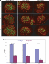
siRNA knock-down in cultured embryonic mouse kidney. siRNA experiments. (A) Photomicrographs showing three representative cultured kidneys stained with laminin (red) and calbindin (green) following either mock transfection (lipofectamine) or transfections with pools of siRNA molecules targeted to Ush2a or Esrrg. No difference is apparent between lipofectamine- and Ush2a-treated kidneys, but kidneys exposed to Esrrg siRNA are smaller and have fewer ductal branch points. (B) Graph showing the quantitative differences in numbers of bud tips (blue) and nephrons (purple). The error bars indicate the 95% confidence intervals and the numbers of kidneys used to generate the data are given above the relevant graph bars.
Figure 5.
Figure 5.
The effect of the Esrrg agonist GSK4716 on cultured embryonic mouse kidney. Effect of exposure to GSK4716. Photomicrographs showing three representative cultured kidneys stained with laminin (red) and calbindin (green) exposed to either none (control) or increasing concentrations (1–10 μm) of the Esrrg agonist GSK4716. At 10 μm concentration, there is a severe developmental anomaly in the exposed embryos with an apparent dose-dependent effect at lower concentrations.
Figure 6.
Figure 6.
Quantitative analysis of Esrrg−/− mouse embryonic kidneys using OPT imaging. Digital sections from OPT scans of paired 17 dpc embryonic kidneys dissected from littermates of Esrrg+/−/Esrrg+/− matings. Eight kidneys from four wild-type embryos (WT), six kidneys from three Esrrg+/− embryos (HET) and six kidneys from three Esrrg−/− embryos (KO) are shown in midline saggital plane defined by analysis of the 3D digital image. The graph shows the range (box), mean (solid line) and error bars (dotted lined) for each genotype group of the luminal volume (right-hand graph) and ratio of lumen to kidney parenchyma for whole kidneys.
Figure 7.
Figure 7.
Pathological features of Esrrg−/− mouse embryonic kidneys. Photomicrographs of sections from wild-type (A, B, C) and Esrrg−/− (D, E, F) littermate 17 dpc embryos. (A) The wild-type can be clearly divided into an outer cortex containing the vast majority of developing glomeruli and an inner medullary region in which a blunt papilla is projecting into a mildly dilated pelvis. (B) The wild-type cortex shows a maturational gradient of nephrogenesis from subcapsular condensation of metanephric mesenchyme around ureteric buds, slightly deeper epithelialization, early morphogenesis (comma and s-shaped bodies), nascent glomeruli at the outer third of the cortex and maturing glomeruli and maturation of convoluted tubules in the deeper cortex. (C) The wild-type medulla, in addition to prominent radiating collecting ducts, contains scattered primitive loops of Henle. Interstitial mesenchymal cells are plump fusiform cells beginning to form parallel arrays. (D) The Esrrg−/− kidney has a marked distortion of the medullary architecture with nearly normal cortical architecture and nephrogenesis. The medulla, instead of forming a normal blunt cone projecting into the pelvis, forms a concave disc beneath the cortex over a widely dilated pelvis. The ureter, however, is not dilated. (E) The cortex shows the same maturational gradient as the wild-type from condensation through tubular maturation. Glomerular maturation appears slightly retarded. (F). The medulla contains radiating collecting ducts, primitive loops of Henle and palisades of interstitial cells but lacks a papilla.
Figure 8.
Figure 8.
Esrrg−/− mouse embryonic kidneys appear normal at 14.5 dpc. Photomicrographs of sections from wild-type (A, B, C), heterozygous (D, E, F) and Esrrg−/− (G, H, I) littermate 14.5 dpc embryos. These sections have been stained with H&E (A, D, G), an antibody that detects phosphorylated histone H3 as a marker of cells undergoing mitosis (pH 3; B, E, H) and an antibody that detects activated caspase 3 as a marker for apoptosis (Caspase; C, F, I). No obvious differences could be detected between the genotypes in any of the staining groups. There were relatively few apoptotic cells in the kidney at this stage and those that were present localized to the ducts (white arrowheads).

References

    1. Hiruma T., Nakamura H. Origin and development of the pronephros in the chick embryo. J. Anat. 2003;203:539–552. doi:10.1046/j.1469-7580.2003.00245.x. - DOI - PMC - PubMed
    1. O'Rahilly R., Müller F. Developmental Stages in Human Embryos. Carnegie Institution of Washington; 1987.
    1. Costantini F. Renal branching morphogenesis: concepts, questions, and recent advances. Differentiation. 2006;74:402–421. doi:10.1111/j.1432-0436.2006.00106.x. - DOI - PubMed
    1. Dressler G.R. The cellular basis of kidney development. Annu. Rev. Cell Dev. Biol. 2006;22:509–529. doi:10.1146/annurev.cellbio.22.010305.104340. - DOI - PubMed
    1. Woolf A.S., Price K.L., Scambler P.J., Winyard P.J. Evolving concepts in human renal dysplasia. J. Am. Soc. Nephrol. 2004;15:998–1007. doi:10.1097/01.ASN.0000113778.06598.6F. - DOI - PubMed

Publication types

Supplementary concepts