. 2011 Mar;39(5):e30.
doi: 10.1093/nar/gkq1258. Epub 2010 Dec 7.

Directed adenovirus evolution using engineered mutator viral polymerases

Affiliations

Directed adenovirus evolution using engineered mutator viral polymerases

Taco G Uil et al. Nucleic Acids Res. 2011 Mar.

Abstract

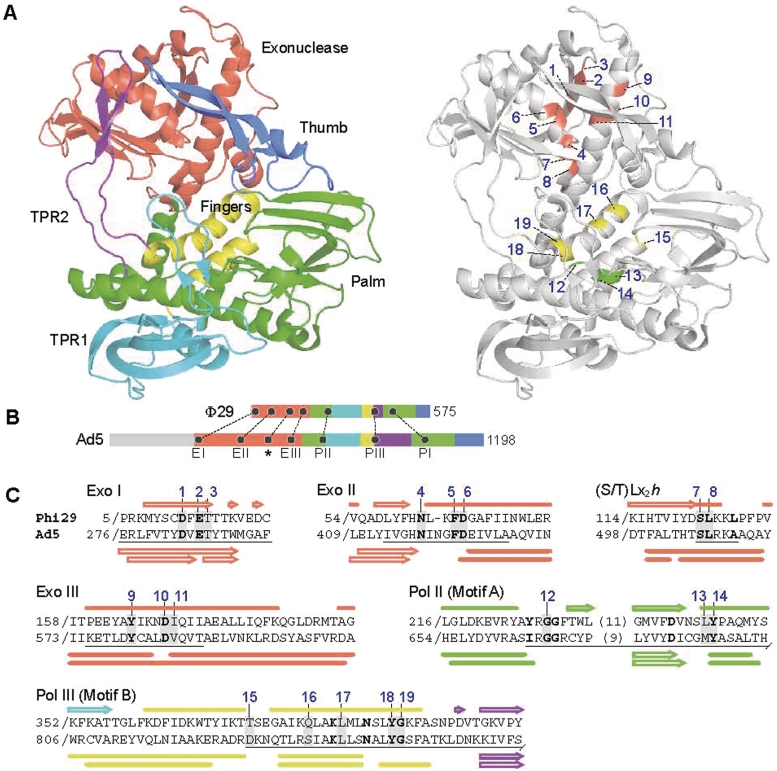
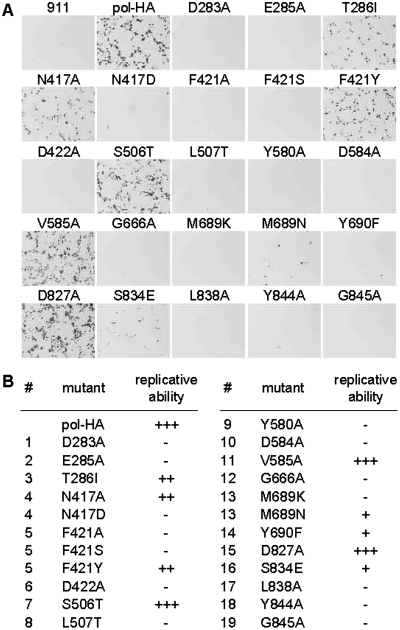
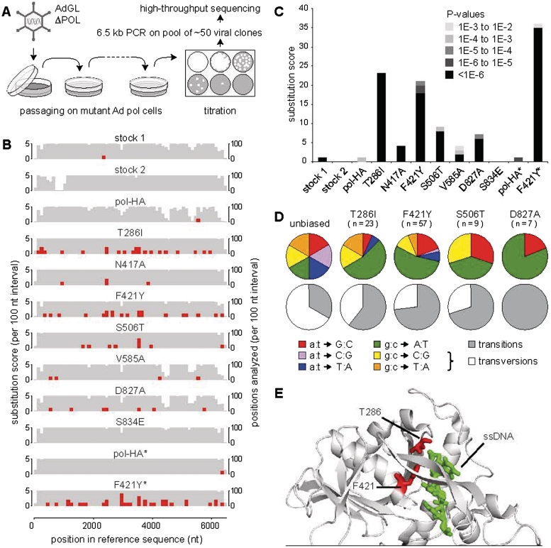
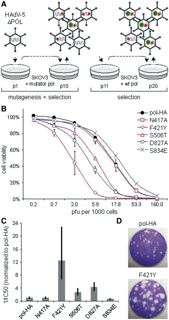
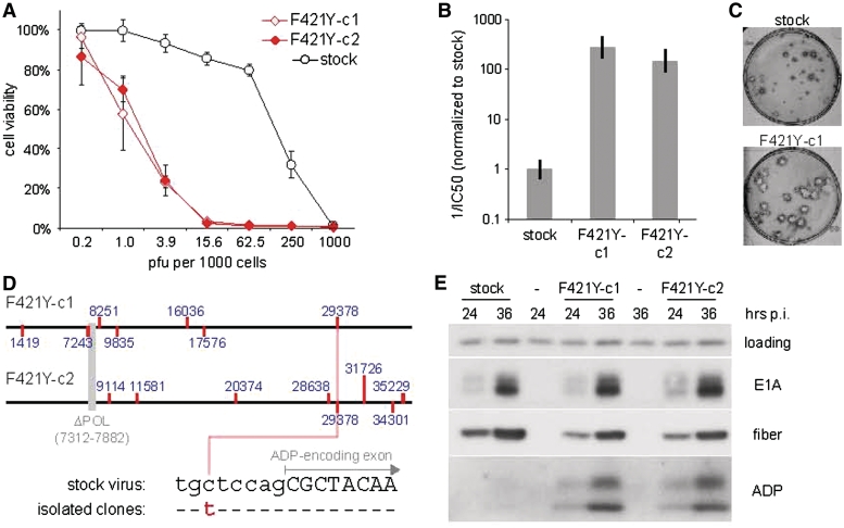
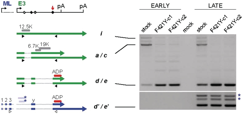
Adenoviruses (Ads) are the most frequently used viruses for oncolytic and gene therapy purposes. Most Ad-based vectors have been generated through rational design. Although this led to significant vector improvements, it is often hampered by an insufficient understanding of Ad's intricate functions and interactions. Here, to evade this issue, we adopted a novel, mutator Ad polymerase-based, 'accelerated-evolution' approach that can serve as general method to generate or optimize adenoviral vectors. First, we site specifically substituted Ad polymerase residues located in either the nucleotide binding pocket or the exonuclease domain. This yielded several polymerase mutants that, while fully supportive of viral replication, increased Ad's intrinsic mutation rate. Mutator activities of these mutants were revealed by performing deep sequencing on pools of replicated viruses. The strongest identified mutators carried replacements of residues implicated in ssDNA binding at the exonuclease active site. Next, we exploited these mutators to generate the genetic diversity required for directed Ad evolution. Using this new forward genetics approach, we isolated viral mutants with improved cytolytic activity. These mutants revealed a common mutation in a splice acceptor site preceding the gene for the adenovirus death protein (ADP). Accordingly, the isolated viruses showed high and untimely expression of ADP, correlating with a severe deregulation of E3 transcript splicing.


Figures

Figure 1.
Figure 1.
Ad pol residues targeted for mutation. Twenty-three single-amino acid substitution mutants of Ad pol were generated by mutation of nineteen selected residues implicated in governing replication fidelity (see also Supplementary Table S1). (A) The respective putative Φ29 homologues of the targeted Ad pol residues are depicted in the context of a Φ29 pol structure (Protein Data Bank code 1XI1) determined by Kamtekar et al. (47). The cartoon representation on the left shows the domain organization of Φ29 pol while the right diagram points out the targeted residues. Protein structure visualizations were by Polyview-3D (http://polyview.cchmc.org/) using image rendering by PyMol. (B) The mutated residues reside in several of the most highly conserved polymerase sequence motifs, the locations of which are shown in linear representations, to scale, of the Φ29 and Ad polymerases. Depicted are the ‘Exo’ motifs I, II and III (59), the (S/T)Lx2h motif (46) (indicated by the asterisk) and the ‘Pol’ regions I, II and III (72), which are also, respectively, known as polymerase motifs C, A and B (73). (C) Sequence alignments of conserved motifs of Ad and Φ29 polymerases. The respective conserved motifs are underlined. Invariable residues are shown in bold face. Blue numbers point to the targeted Ad pol residues and their respective putative Φ29 homologues. Secondary structures known for Φ29 (47) and those predicted for Ad pol are, respectively, shown above and below the alignments. Closed bars indicate α-helices while open arrows represent β-strands. The upper and lower predictions for Ad pol were, respectively, by PredictProtein (http://www.predictprotein.org) and SABLE (http://sable.cchmc.org).
Figure 2.
Figure 2.
Ability of candidate mutator Ad polymerases to support full-fledged Ad replication. (A) A trans-complementation system (see Supplementary Figure S1 for its components) was employed to assess the ability of the various Ad pol mutants to support Ad replication. Control 911 cells or 911 cell populations stably expressing the respective Ad pol variants were infected with a polymerase-defective Ad vector (AdGLΔPOL). The mutants’ abilities to trans-complement AdGLΔPOL were assessed by monitoring vector-encoded reporter gene expression. The images show GFP fluorescence (inverted grayscale) at 2 days post-infection. ‘pol-HA’ stands for HA-tagged Ad pol, which is parental to the other Ad pol variants. The exposure time was the same for all pictures. Not discernible under this setting was the very weak GFP signal in infected cells that do not trans-complement the polymerase defect (e.g. 911 cells). (B) Summary of the abilities of the Ad pol mutants to complement the polymerase-defective vector. The numbers in the columns denoted by a pound symbol (#) correspond to the residue numbers assigned in Figure 1. The depicted complementation classes (−, +, ++ and +++) were arbitrarily assigned to reflect the differential patterns of complementation abilities observed for the mutants in several independent experiments (e.g. Figure 2A).
Figure 3.
Figure 3.
Identification of mutator Ad polymerases. (A) Mutation-accumulation and deep sequencing scheme. A polymerase-defective vector, AdGLΔPOL, was subjected to 10 serial infection rounds on candidate mutator Ad pol-expressing cell populations. For each passaged virus population, the potential mutation load buildup was assessed by deep sequencing (using a MPS technology) of a 6.5-kb virus DNA fragment obtained from a ‘50-clone’ virus pool. After mapping of the sequence reads to the reference sequence, single-base substitutions were scored using a minor variant frequency cutoff value of 0.25% and a minimal sequencing depth requirement of 1200, both of which conditions were to be met for both the forwardly and reversely mapped read distributions. See Supplementary Figure S3 for details on mappings and minor variant detection. (B) Substitution score distributions over the length of the sequenced fragment. The grey background columns indicate, per 100-nt interval, the number of positions included in the analysis (i.e. the number of positions for which the minimal sequencing depth requirement was met). The red columns display the single-base substitutions scores. ‘Stock 1’ and ‘stock 2’ represent two independently prepared 50-clone pools of non-passaged viruses. Sample names marked with an asterisk (*) indicate the use of an alternative pooling method to obtain the ‘50-clone’ pools. See text for details. (C) Total number of substitutions found per analyzed pool. For a given substitution, the assigned P-value range (shade of gray) relates to the higher of the two P-values estimated for the forward and reverse occurrence of that substitution. See the statistics section for P-value estimations. Further, see Supplementary Table S2 for the substitution scores expressed relative to the amount of DNA analyzed. (D) Base substitution spectra of the identified mutator Ad polymerases. The relative frequencies of the different types of substitutions were corrected for the GC content (of ∼60%) of the analyzed sequence. (E) The Φ29 homologues of Ad Pol residues T286 and F421 have previously been shown to entertain direct contacts with single-stranded DNA (ssDNA) (47). The concerned residues are depicted, with side chains, in a cartoon representation of Φ29 pol, additionally displaying the cocrystallized ssDNA bound in the exonuclease active site. See Figure 1 for protein structure and visualization.
Figure 4.
Figure 4.
Directed evolution of Ad using mutator Ad polymerases. (A) Conceptual directed evolution scheme to achieve enhanced tumor cell killing. A HAdV-5-based virus, which otherwise from its polymerase disrupting deletion is fully wildtype in sequence, is subjected to iterative rounds of infection on tumor cell populations. Initial virus passaging on mutator Ad pol expressing cells results in genetically diverse pools from which viruses with an acquired growth advantage are enriched for in later passages. Red diamonds represent mutations. Green circles indicate an acquired selective advantage. (B) Virus populations bioselected using the indicated Ad pol variants were analyzed for cytotoxicity on their target cells. SKOV-3 cells were infected at the indicated MOI’s and cell viabilities were assessed by WST-1 assay. Exonuclease domain mutants are shown in red, fingers domain mutants in blue. (C) A 12-fold decrease in IC50 was observed for the virus population bioselected using Ad pol mutator F421Y. The inversed IC50 values were calculated from the cytotoxicity assay above and normalized to that of the pol-HA-bioselected pool. Error bars indicate 95% confidence intervals. (D) A subpopulation of the F421Y-bioselected viruses forms large plaques on SKOV-3 cell monolayers. Cells were grown under an agarose layer and stained with crystal violet.
Figure 5.
Figure 5.
Characterization of isolated bioselected viral clones F421Y-c1 and F421Y-c2. Two clones with a large plaque phenotype were isolated from the F421Y-bioselected virus population and further characterized. Cell killing ability on SKOV-3 cells was assessed by a cytotoxicity (A and B) and a plaque size (C) assay. Error bars for the inversed IC50 columns indicate 95% confidence intervals. For the plaque size assay, cells were grown under an agarose layer and were visualized by immunohistochemistry for Ad fiber protein expression. See Supplementary Figure S4 for cell killing abilities on other (tumor) cell lines. (D) Sequencing of the genomes of the two clones revealed a shared mutation located within the splice acceptor site of the ADP-encoding exon. Mutations are indicated by red ticks. Blue numbers correspond to the respective positions in the wildtype HAdV-5 genome (accession no. AC_000008). See Supplementary Tables S3 and S4 for all detected mutations and their predicted consequences for primary protein structures. (E) The bioselected clones show highly increased levels of ADP expression. SKOV-3 cells were harvested for protein immunoblot analysis at 24 and 36 h after their infection. Blots were immunologically probed for Ad proteins E1A, fiber and ADP.
Figure 6.
Figure 6.
Viral clones F421Y-c1 and F421Y-c2 show a deregulated E3 splicing pattern strongly favoring the production of ADP-encoding mRNAs. RT–PCRs were performed with primer sets discriminating between E3 and ML promoter-derived ADP-encoding mRNAs. These primer sets, respectively, indicated by closed and open triangles, have a common reverse primer (targeting the ADP coding sequence) but consist of different forward primers (targeting the first E3 exon and the 3rd leader of the ML tripartite leader sequence). Depicted are all mRNA species recognized by these primer sets [according to Ad2 and Ad5 transcritption maps (42,48,49)]. E3 mRNAs (i.e. a, c, d, e and i) are shown in green while ML mRNAs (i.e. d’ and e’) are depicted in blue. Within the E3 transcription unit, the splice donor and acceptor sites involved with splicing of the shown mRNAs are, respectively, depicted by open and closed diamonds. The red arrow points to the splice site found to be mutated in the bioselected clones. Further depicted are the relevant polyadenylation signals (pA), E3 proteins (12.5K, 6.7K, 19K and ADP) and leader sequences of the ML transcription unit (1, 2, 3 and y). The RT–PCR results show highly increased levels of E3- but not ML-derived ADP mRNAs. Furthermore, the altered alternative splicing of E3 transcripts was found to go at the expense of mRNA species a/c and i. The two asterisks next to the gel-image denote newly identified minority ML-derived ADP mRNAs. These variants presumably carry instead of the y-leader sequence one of two longer ‘x-leader’ exons (also denoted by asterisks) that would overlap with the first E3 exon. For control RT–PCRs on cellular β-actin and viral E1A mRNAs see Supplementary Figure S5.

Similar articles

Cited by

References

    1. Parato KA, Senger D, Forsyth PA, Bell JC. Recent progress in the battle between oncolytic viruses and tumours. Nat. Rev. Cancer. 2005;5:965–976. - PubMed
    1. Alemany R. Designing adenoviral vectors for tumor-specific targeting. Methods Mol. Biol. 2009;542:57–74. - PubMed
    1. Toth K, Dhar D, Wold WS. Oncolytic (replication-competent) adenoviruses as anticancer agents. Expert. Opin. Biol. Ther. 2010;10:353–368. - PubMed
    1. DeWeese TL, van der PH, Li S, Mikhak B, Drew R, Goemann M, Hamper U, DeJong R, Detorie N, Rodriguez R, et al. A phase I trial of CV706, a replication-competent, PSA selective oncolytic adenovirus, for the treatment of locally recurrent prostate cancer following radiation therapy. Cancer Res. 2001;61:7464–7472. - PubMed
    1. Khuri FR, Nemunaitis J, Ganly I, Arseneau J, Tannock IF, Romel L, Gore M, Ironside J, MacDougall RH, Heise C, et al. a controlled trial of intratumoral ONYX-015, a selectively-replicating adenovirus, in combination with cisplatin and 5-fluorouracil in patients with recurrent head and neck cancer. Nat. Med. 2000;6:879–885. - PubMed

Publication types

Substances