. 2011 Jan;138(1):139-48.
doi: 10.1242/dev.054239.

Retinoic acid stimulates myocardial expansion by induction of hepatic erythropoietin which activates epicardial Igf2

Affiliations

Retinoic acid stimulates myocardial expansion by induction of hepatic erythropoietin which activates epicardial Igf2

Thomas Brade et al. Development. 2011 Jan.

Abstract

Epicardial signaling and Rxra are required for expansion of the ventricular myocardial compact zone. Here, we examine Raldh2(-/-) and Rxra(-/-) mouse embryos to investigate the role of retinoic acid (RA) signaling in this developmental process. The heart phenotypes of Raldh2 and Rxra mutants are very similar and are characterized by a prominent defect in ventricular compact zone growth. Although RA activity is completely lost in Raldh2(-/-) epicardium and the adjacent myocardium, RA activity is not lost in Rxra(-/-) hearts, suggesting that RA signaling in the epicardium/myocardium is not required for myocardial compact zone formation. We explored the possibility that RA-mediated target gene transcription in non-cardiac tissues is required for this process. We found that hepatic expression of erythropoietin (EPO), a secreted factor implicated in myocardial expansion, is dependent on both Raldh2 and Rxra. Chromatin immunoprecipitation studies support Epo as a direct target of RA signaling in embryonic liver. Treatment of an epicardial cell line with EPO, but not RA, upregulates Igf2. Furthermore, both Raldh2(-/-) and Rxra(-/-) hearts exhibit downregulation of Igf2 mRNA in the epicardium. EPO treatment of cultured Raldh2(-/-) hearts restores epicardial Igf2 expression and rescues ventricular cardiomyocyte proliferation. We propose a new model for the mechanism of RA-mediated myocardial expansion in which RA directly induces hepatic Epo resulting in activation of epicardial Igf2 that stimulates compact zone growth. This RA-EPO-IGF2 signaling axis coordinates liver hematopoiesis with heart development.


Figures

Fig. 1.
Fig. 1.
Comparison of heart phenotype in rescued Raldh2–/– and Rxra–/– mouse embryos. (A-F) Sections of Hematoxylin and Eosin-stained E13.5 hearts from wild-type (A,D), germline Rxra–/– (B,E) and rescued Raldh2–/– (C,F) embryos. Representative heart sections are shown at 40× magnification in A-C and detailed photographs of the compact zone at 400× magnification in D-F. Both mutants exhibit a severe reduction in the size of the compact zone myocardium (CZ) whereas the trabecular myocardium (Tr) is only mildly affected; the black bar indicates the thickness of the compact zone. Similar results were observed for all mutants analyzed (n=3 for each genotype).
Fig. 2.
Fig. 2.
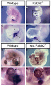
Detection of cardiac RA signaling in rescued Raldh2–/– and Rxra–/– mouse embryos. (A-L) RARE-lacZ staining of wild-type (A,D,G,J), germline Rxra–/– (B,E,H,K) and rescued Raldh2–/– (C,F,I,L) whole hearts and sections at E10.5 (A-F) and E12.5 (G-L). Similar results were observed for all mutants analyzed (n=3 for each genotype and stage). RA signaling is not altered in germline Rxra–/– hearts at E10.5 and E12.5 relative to wild-type hearts, whereas loss of Raldh2 leads to severe impairment of RA signaling in cardiac tissues at both stages, including loss of epicardial RA signaling at E12.5 (compare J,K,L). a, atrium; E, epicardium; ot, outflow tract; v, ventricle.
Fig. 3.
Fig. 3.
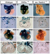
Epicardium develops in rescued Raldh2–/– mouse embryos. (A-D) At E9.5, detection of Tbx18 mRNA indicates that the proepicardial organ is present in both wild-type (A,C) and unrescued Raldh2–/– (B,D) embryos. C and D are higher magnifications of the boxed areas in A and B, respectively. (E-H) E10.5 hearts of both wild-type and rescued Raldh2–/– mutants exhibit Tbx18 mRNA expression demonstrating that the epicardium has developed in the mutant. G and H are higher magnifications of the boxed areas in E and F, respectively. Similar Tbx18 detection was observed for all mutants analyzed (n=3 for both stages). A, atrium; E, epicardium; H, heart; p, proepicardial organ; V, ventricle.
Fig. 4.
Fig. 4.
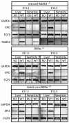
Analysis of Epo and Fgf9 expression in hepatic and ventricular mouse tissues. Semi-quantitative RT-PCR shows that Epo mRNA is reduced in the liver of rescued Raldh2–/– embryos at E11.5 and E12.5 relative to wild type, whereas Epo expression is not detectable in ventricular tissues; as a control, rescued Raldh2–/– embryos exhibit a loss of Raldh2 mRNA. Epo mRNA is also reduced in livers from germline Rxra–/– embryos and Gata5-Cre x Rxra–/– conditional mutant embryos. Expression of Fgf9 mRNA is not altered in any of the analyzed tissues and mutants relative to wild type. GAPDH, glyceraldehyde 3-phosphate dehydrogenase mRNA control.
Fig. 5.
Fig. 5.
Recruitment of RA receptors to the Epo 3′ enhancer in mouse embryonic liver. (A) Schematic representation of the 3′ enhancer region of mouse Epo showing the location of a DR2 RARE (direct repeat with 2 bp spacer) and PCR primers used for chromatin immunoprecipitation (ChIP) analysis. (B) E13.5 liver ChIP results demonstrating robust binding of all three RARs detected with primers flanking the Epo RARE; no signal was obtained with IgG control or non-specific primers located several kb upstream from the RARE. M, DNA ladder; S, RARE specific primers; NS, specific primers.
Fig. 6.
Fig. 6.
RA controls cardiac IGF2 expression indirectly through EPO. (A) Treatment of epicardial MEC-1 cell line with EPO (10 ng/ml) significantly induced Igf2 mRNA compared with cells grown under serum-free conditions (SF), whereas RA treatment (1 μM) had no effect; expression based on RT-PCR analysis was normalized to β-actin mRNA. Data are mean ± s.d. (B) Igf2 mRNA was detected by in situ hybridization in E12.5 mouse hearts. Rescued Raldh2–/– and Rxra–/– hearts exhibit a reduction in epicardial Igf2 mRNA detection compared with wild type. The bar represents the thickness of the compact zone. Similar results were observed for all mutants analyzed (n=3 for each genotype). cz, compact zone myocardium; ep, epicardium.
Fig. 7.
Fig. 7.
Rescue of epicardial Igf2 expression. (A-D) Wild-type and Raldh2–/– mouse hearts were cultured in the absence or presence of EPO, then sections were analyzed for Igf2 expression by in situ hybridization. Wild-type hearts (A,B) exhibited Igf2 mRNA with (Epo) or without (Con) added EPO. A control (con) Raldh2–/– heart (C) displays no epicardial Igf2 mRNA, but EPO treatment rescues epicardial Igf2 expression in an Raldh2–/– heart (D; n=3 for each genotype). ep, epicardium.
Fig. 8
Fig. 8
Cardiomyocyte proliferation in heart organ culture is rescued by EPO treatment. Hearts dissected from E11.5 wild-type (WT) or Raldh2–/– mutant mice were cultured for 18 hours in the absence of EPO (control) or with EPO (+EPO), then examined immunohistochemically for cardiomyocytes (MF20; red), dividing cells (Ki67; green), and nuclei (DAPI; blue). Control or EPO-treated wild-type hearts exhibited significantly more proliferating cardiomyocytes (MF20+, Ki67+) compared with control Raldh2–/– hearts, but EPO treatment of Raldh2–/– hearts restored the cardiomyocyte proliferation rate to wild-type levels (n=2 for each genotype). Data are mean ± s.d.
Fig. 9.
Fig. 9.
RA-EPO-IGF2 signaling axis from liver to heart. We propose a model for control of myocardial compact zone formation that involves a sequence of three signaling pathways extending from liver to heart: (1) RA signaling generated by hepatic RALDH2 and RXRα induces hepatic Epo; (2) EPO secreted by the liver results in epicardial EPO signaling needed to induce Igf2; (3) IGF2 secreted by the epicardium results in myocardial IGF2 signaling needed to stimulate ventricular myocardial growth. INSr/IGF1r represents the receptors for IGF2.

References

    1. Asahina K., Tsai S. Y., Li P., Ishii M., Maxson R. E., Jr, Sucov H. M., Tsukamoto H. (2009). Mesenchymal origin of hepatic stellate cells, submesothelial cells, and perivascular mesenchymal cells during mouse liver development. Hepatology 49, 998-1011 - PMC - PubMed
    1. Azambuja A. P., Portillo-Sanchez V., Rodrigues M. V., Omae S. V., Schechtman D., Strauss B. E., Costanzi-Strauss E., Krieger J. E., Perez-Pomares J. M., Xavier-Neto J. (2010). Retinoic Acid and VEGF delay smooth muscle relative to endothelial differentiation to coordinate inner and outer coronary vessel wall morphogenesis. Circ. Res. 107, 204-216 - PubMed
    1. Bussen M., Petry M., Schuster-Gossler K., Leitges M., Gossler A., Kispert A. (2004). The T-box transcription factor Tbx18 maintains the separation of anterior and posterior somite compartments. Genes Dev. 18, 1209-1221 - PMC - PubMed
    1. Calleja C., Messaddeq N., Chapellier B., Yang H., Krezel W., Li M., Metzger D., Mascrez B., Ohta K., Kagechika H., et al. (2006). Genetic and pharmacological evidence that a retinoic acid cannot be the RXR-activating ligand in mouse epidermis keratinocytes. Genes Dev. 20, 1525-1538 - PMC - PubMed
    1. Chen J., Kubalak S. W., Chien K. R. (1998). Ventricular muscle-restricted targeting of the RXRα gene reveals a non-cell-autonomous requirement in cardiac chamber morphogenesis. Development 125, 1943-1949 - PubMed

Publication types

MeSH terms