. 2011 Feb 15;89(3):671-9.
doi: 10.1093/cvr/cvq346. Epub 2010 Dec 7.

Soluble FLT1 sensitizes endothelial cells to inflammatory cytokines by antagonizing VEGF receptor-mediated signalling

Affiliations

Soluble FLT1 sensitizes endothelial cells to inflammatory cytokines by antagonizing VEGF receptor-mediated signalling

Tereza Cindrova-Davies et al. Cardiovasc Res. .

Abstract

Aims: Pre-eclampsia affects 5-7% of pregnancies, and is a major cause of maternal and foetal death. Elevated serum levels of placentally derived splice variants of the vascular endothelial growth factor (VEGF) receptor, soluble fms-like tyrosine kinase-1 (sFLT1), are strongly implicated in the pathogenesis but, as yet, no underlying mechanism has been described. An excessive inflammatory-like response is thought to contribute to the maternal endothelial cell dysfunction that characterizes pre-eclampsia. We hypothesized that sFLT1 antagonizes autocrine VEGF-A signalling, rendering endothelial cells more sensitive to pro-inflammatory factors also released by the placenta. We tested this by manipulating VEGF receptor signalling and treating endothelial cells with low doses of tumour necrosis factor-α (TNF-α).

Methods and results: Application of recombinant sFLT1 alone did not activate human umbilical vein endothelial cells (HUVECs). However, antagonizing the autocrine actions of endothelial VEGF-A and/or placenta growth factor (PlGF) by pre-incubation with recombinant sFLT1, anti-FLT1, anti-VEGF receptor 2 (KDR), anti-VEGF-A, VEGF receptor tyrosine kinase inhibitor SU5614, or knocking-down FLT1 or KDR transcripts rendered cells more sensitive to low doses of TNF-α. Each treatment increased activation, as measured by increases in endothelial intercellular adhesion molecule 1 (ICAM1), vascular cell adhesion molecule 1 (VCAM1), endothelin 1 (ET-1), von Willebrand factor (vWF), and leucocyte adhesion, and led to reduction in AKT Ser⁴⁷³ and endothelial nitric oxide synthase (eNOS) Ser¹¹⁷⁷ phosphorylation.

Conclusions: Our data describe a mechanism by which sFLT1 sensitizes endothelial cells to pro-inflammatory factors, providing an explanation for how placental stress may precipitate the pre-eclamptic syndrome.


Figures
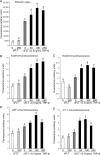
Figure 1
Figure 1
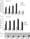
Pre-treatment of HUVECs with sFLT1 significantly increases HL60 leucocyte adhesion, and expression of ICAM1, VCAM1, vWF, and ET-1 upon TNF-α treatment. HUVECs were pre-treated with/without sFLT1 (50–250 ng/mL) for 24 h and subsequently treated with TNF-α for 6 h. Fluorescently labelled HL60 leucocytes were added in the final hour of TNF-α incubation and fluorescence was detected (A). Fixed HUVECs were immunostained against ICAM1 (B), VCAM1 (C), vWF (D), and ET-1 (E), and fluorescence was quantified. Experiments were repeated at least three times. Letters indicate groups significantly different using the PLSD test with P< 0.05.
Figure 2
Figure 2
HL60 leucocyte adhesion upon TNF-α treatment is significantly increased in HUVECs transfected with siKDR or siFLT1 or treated with neutralizing antibodies against KDR, FLT1, or VEGF-A or with the VEGF receptor kinase inhibitor SU5614. HUVECs were transfected with siKDR, siFLT1, siTNFR-SF1A, or siICAM for 24 h (A), pre-treated with/without sFLT and/or anti-KDR, anti-FLT1 or anti-VEGF-A (B) for 24 h and then treated with TNF-α for 6 h. Fluorescently labelled HL60 leucocytes were added in the final hour of TNF-α incubation and fluorescence was detected. Western blots were run to detect ICAM1 expression (C). These graphs represent one experimental treatment. Experiments were repeated at least three times. Letters indicate groups significantly different using the PLSD test with P< 0.05. F100—sFLT1 100 ng/mL; T0.5—TNF-α 0.5 ng/mL.
Figure 3
Figure 3
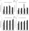
Treatment of HUVECs with neutralizing antibodies against KDR, FLT1, or VEGF-A or with the VEGF receptor kinase inhibitor SU5614 increases the expression of ICAM1, VCAM1, vWF, and ET-1 upon TNF-α treatment. HUVECs were pre-treated with/without sFLT for 24 h, followed by a 30-min pre-treatment with/without anti-KDR, anti-FLT1, or anti-VEGF-A and then treated with TNF-α for 6 h. Fixed HUVECs were immunostained against ICAM1 (A), VCAM1 (B), vWF (C), or ET-1(D) and immunofluorescence was quantified. Each graph represents one experimental treatment. Experiments were repeated at least three times. Letters indicate groups significantly different using the PLSD test with P< 0.05. F100—sFLT1 100 ng/mL; T0.5—TNF-α 0.5 ng/mL.
Figure 4
Figure 4
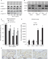
The role of the AKT pathway in sFLT1 signalling. Lysates from HUVECs pre-treated with/without sFLT1 (10–200 ng/mL) for 24 h and subsequently treated with TNF-α for 6 h were immunoblotted with antibodies against P-AKTSer473, AKT, P-eNOSSer1177, and eNOS (A) and the signal from three experiments was quantified (C). HUVECs transfected with siAKT1 or siAKT3 for 24 h and then treated with TNF-α for 6 h were either immunoblotted with antibodies against AKT1, AKT3, and ICAM1 (B) or incubated with fluorescently labelled HL60 leucocytes in the final hour of TNF-α incubation and fluorescence was detected (D). Each experiment was repeated at least three times. Letters indicate groups significantly different using the PLSD test with P< 0.05. In Figure 4C, lower-case letters compare the signal of P-AKTSer473 and capital letters the signal of P-eNOSSer1177. Columns labelled with the same letter do not differ significantly. (E) Immunostaining of caesarean delivered placentas for AKT1, AKT2, and AKT3 localized these to trophoblast and endothelial cells (AKT1), trophoblast only (AKT2), or endothelial cells only (AKT3).
Figure 5
Figure 5
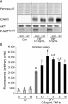
Inhibition of the AKT pathway potentiates the action of TNF-α. HUVECs pre-treated with LY294002 (LY; 0.1–10 μM) and then treated with TNF-α for 6 h were either harvested for western blotting and probed with antibodies against P-AKTSer473 or ICAM1 (A) or incubated with fluorescently labelled HL60 leucocytes in the final hour of TNF-α incubation and fluorescence was detected (B). Each experiment was repeated at least three times. Letters indicate groups significantly different using the PLSD test with P< 0.05.

References

    1. Bethesda M WHO. Make Every Mother and Child Count. National Institutes of Health: WHO; 2005. Global burden of disease for the year 2001 by World Bank region, for use in disease control priorities in developing countries. WHRGWHOW, 2004. http://www.who.int/whr/2005/chapter3/en/index2.html .
    1. Redman CW, Sargent IL. Latest advances in understanding preeclampsia. Science. 2005;308:1592–1594. doi:10.1126/science.1111726. - DOI - PubMed
    1. Roberts JM. Endothelial dysfunction in preeclampsia. Semin Reprod Endocrinol. 1998;16:5–15. doi:10.1055/s-2007-1016248. - DOI - PubMed
    1. Myatt L, Webster RP. Vascular biology of preeclampsia. J Thromb Haemost. 2009;7:375–384. doi:10.1111/j.1538-7836.2008.03259.x. - DOI - PubMed
    1. Maynard SE, Min JY, Merchan J, Lim KH, Li J, Mondal S, et al. Excess placental soluble fms-like tyrosine kinase 1 (sFlt1) may contribute to endothelial dysfunction, hypertension, and proteinuria in preeclampsia. J Clin Invest. 2003;111:649–658. - PMC - PubMed

Publication types

MeSH terms

Substances