. 2011 Mar;10(3):M110.003590.
doi: 10.1074/mcp.M110.003590. Epub 2010 Dec 7.

Mass spectrometric analysis of lysine ubiquitylation reveals promiscuity at site level

Affiliations

Mass spectrometric analysis of lysine ubiquitylation reveals promiscuity at site level

Jannie M R Danielsen et al. Mol Cell Proteomics. 2011 Mar.

Abstract

The covalent attachment of ubiquitin to proteins regulates numerous processes in eukaryotic cells. Here we report the identification of 753 unique lysine ubiquitylation sites on 471 proteins using higher-energy collisional dissociation on the LTQ Orbitrap Velos. In total 5756 putative ubiquitin substrates were identified. Lysine residues targeted by the ubiquitin-ligase system show no unique sequence feature. Surface accessible lysine residues located in ordered secondary regions, surrounded by smaller and positively charged amino acids are preferred sites of ubiquitylation. Lysine ubiquitylation shows promiscuity at the site level, as evidenced by low evolutionary conservation of ubiquitylation sites across eukaryotic species. Among lysine modifications a significant overlap (20%) between ubiquitylation and acetylation at site level highlights extensive competitive crosstalk among these modifications. This site-specific crosstalk is not prevalent among cell cycle ubiquitylations. Between SUMOylation and ubiquitylation the preferred interaction is through mixed-chain conjugation. Overall these data provide novel insights into the site-specific selection and regulatory function of lysine ubiquitylation.


Figures

Fig. 1.
Fig. 1.
Characterization of a stable cell line expressing ectopic Strep-HA-ubiquitin. A, Whole-cell extracts (WCE) of U2OS cells stably expressing Strep-HA-tagged ubiquitin were subjected to Strep-Tactin Sepharose pull down and analyzed by immunoblotting with HA antibody. B, Lysates from U2OS/Strep-HA-ubiquitin cells were subjected to immunoblotting with indicated antibodies. C, U2OS/Strep-HA-ubiquitin cells grown on coverslips were fixed and immunostained with HA antibody. Scale bar, 10 μm. D, U2OS cells or U2OS/Strep-HA-ubiquitin were mock-treated or incubated in the presence of TNF-α, and harvested 1 h later. RIP1 ubiquitylation was analyzed by immunoblotting of Strep-Tactin Sepharose pull downs from cell extracts with RIP1 antibody. E, U2OS/Strep-HA-ubiquitin cells were processed as in (A), resolved on SDS-PAGE and Coomassie-stained. The lane containing Strep-protein complexes was divided into 20 slices and processed for mass spectrometric analysis of site-specific ubiquitylation.
Fig. 2.
Fig. 2.
A, Overlap of ubiquitylation sites from three analyzed experiments. Experiments one and two were biological U2OS replicates, whereas experiment three was enrichment analysis of HEK293T cells. B, Overlap of identified ubiquitylation substrates from same three experiments. C, Normalized abundance measurement of poly-ubiquitin chain linkages on ubiquitin by MS. Data follows previous results reported in the literature. D, Lysates from U2OS cells co-transfected with indicated combinations of Myc-ubiquitin and FLAG-Cbx1 constructs were subjected to FLAG immunoprecipitation followed by immunoblotting with Myc antibody to visualize Cbx1 ubiquitylation.
Fig. 3.
Fig. 3.
A, Sequence logo plots represent normalized amino acid frequencies for ±6 amino acids from the lysine ubiquitylation site. No significantly enriched motifs were detected. B, Amino acid property plot representing clustered amino acid groups for a combined ±5 amino acids from the lysine ubiquitylation site. Green bars represent Alanine and Glycine residues, blue bars represent charged residues Lysine and Arginine whereas yellow represent Cysteine. C, Distribution of average lysines and ubiquitylated lysines in structured and unstructured regions of the proteins. A significant enrichment of ubiquitylation sites residing in helical structures is observed. D, Accessibility distribution of average lysines and ubiquitylated lysines. A small but significant preference for accessibility by the ubiquitin system is observed.
Fig. 4.
Fig. 4.
A, Conservation analysis of average lysines and ubiquitylated lysines across eukaryotic species. Ubiquitylated lysines show a slightly higher conservation compared with average lysines (6% and 3% respectively, of lysines being conserved in more than 50% of eukaryotic species). B, Distribution of N-terminal amino acids belonging to (i) 471 proteins identified as being lysine ubiquitylated (blue bars), (ii) all 5.600 putative ubiquitylated proteins identified in entire experiment (red bars) and (iii) all proteins in the human database (green bars). A significant enrichment for N-terminal Serine and Alanine residues is observed among ubiquitylated proteins.
Fig. 5.
Fig. 5.
A, Distribution of site-specific overlap between lysine ubiquitylation and acetylation. Blue bars represent overlap by random, red bars represent observed overlap. B, Identified lysine ubiquitylations belonging to cell cycle proteins determined according to GOBP. Red box represents proteins belonging to M-phase, whereas blue box represents proteins belonging to S-phase. Red balls represent novel ubiquitylation sites identified, yellow balls are known sites not identified in this study, whereas yellow/red balls are previously identified ubiquitylations sites confirmed in this study. C, Normalized abundance measurement of ubiquitin chain linkages upon SUMO2 (mixed chain conjugation). The majority of SUMO2 ubiquitylation occurs through K11 followed by K7, K42, K33, K5, and K21.
Fig. 6.
Fig. 6.
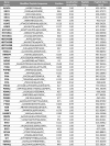
Table of 45 nuclear lysine residues identified as being both ubiquitylated and acetylated. Proteins to which the modified sites belong are listed with gene name. Localization probability of the identified ubiquitylation site, as well as charge state and measured mass (m/z) of the modified peptide are listed.

References

    1. Yang X. J. (2005) Multisite protein modification and intramolecular signaling. Oncogene 24, 1653–1662 - PubMed
    1. Mann M., Jensen O. N. (2003) Proteomic analysis of post-translational modifications. Nat. Biotechnol. 21, 255–261 - PubMed
    1. Ciechanover A. (1994) The ubiquitin-proteasome proteolytic pathway. Cell 79, 13–21 - PubMed
    1. Scheffner M., Nuber U., Huibregtse J. M. (1995) Protein ubiquitination involving an E1-E2-E3 enzyme ubiquitin thioester cascade. Nature 373, 81–83 - PubMed
    1. Hussain S., Zhang Y., Galardy P. J. (2009) DUBs and cancer: the role of deubiquitinating enzymes as oncogenes, non-oncogenes and tumor suppressors. Cell Cycle 8, 1688–1697 - PubMed

Publication types

LinkOut - more resources