. 2011 Mar;31(3):795-806.
doi: 10.1038/jcbfm.2010.204. Epub 2010 Dec 8.

In vivo 3D morphology of astrocyte-vasculature interactions in the somatosensory cortex: implications for neurovascular coupling

Affiliations

In vivo 3D morphology of astrocyte-vasculature interactions in the somatosensory cortex: implications for neurovascular coupling

Addason F H McCaslin et al. J Cereb Blood Flow Metab. 2011 Mar.

Abstract

Astrocytes are increasingly believed to play an important role in neurovascular coupling. Recent in vivo studies have shown that intracellular calcium levels in astrocytes correlate with reactivity in adjacent diving arterioles. However, the hemodynamic response to stimulation involves a complex orchestration of vessel dilations and constrictions that spread rapidly over wide distances. In this work, we study the three-dimensional cytoarchitecture of astrocytes and their interrelations with blood vessels down through layer IV of the mouse somatosensory cortex using in vivo two-photon microscopy. Vessels and astrocytes were visualized through intravenous dextran-conjugated fluorescein and cortically applied sulforhodamine 101 (SR101), respectively. In addition to exploring astrocyte density, vascular proximity, and microvascular density, we found that sheathing of subpial vessels by astrocyte processes was continuous along all capillaries, arterioles, and veins, comprising a highly interconnected pathway through which signals could feasibly be relayed over long distances via gap junctions. An inner SR101-positive sheath noted along pial and diving arterioles was determined to be nonastrocytic, and appears to represent selective SR101 staining of arterial endothelial cells. Our findings underscore the intimate relationship between astrocytes and all cortical blood vessels, and suggest that astrocytes could influence neurovascular regulation at a range of sites, including the capillary bed and pial arterioles.


Figures
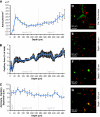
Figure 1
Figure 1
Astrocyte density, capillary density, and astrocyte–vessel separation. (A) Astrocyte density as a function of cortical depth (excluding the glia limitans superficialis between 0 and 20 μm). Cell density was sampled in 20-μm increments. (B) Capillary density as a function of cortical depth, sampled in 2-μm increments. (C) The average separation between astrocytes and capillaries as a function of cortical depth sampled in 20-μm increments. All error bars show s.e.m. Layer I to IV positions are indicated in accordance with Altamura et al (2007), although it should be noted that these boundaries are simply guidelines, and that Tsai et al (2009) found that layer I may start at depths of up to 100 μm in unprocessed tissue. (D to G) Representative images from specific cortical depths. Red=SR101 labeling, green=intravascular dextran-conjugated fluorescein. (Panel D) Image of the glia limitans at the cortical surface. Pial veins are visible with sub-pial perivascular SR101 sheathing merging into the numerous cell bodies of the glia limitans. (Panel E) Dense astrocyte cell bodies 50 μm below the surface of the brain. (Panel F) Sparse astrocyte cell bodies at a depth of 150 μm. Neuron cell bodies are visible as dark, unstained areas. (Panel G) Astrocyte density recovers approaching 450 μm. Capillary density is also elevated. This full stack is provided as a movie and 3D rendering in Supplementary data. SR101, sulforhodamine 101.
Figure 2
Figure 2
Perivascular sheathing morphology. (A) Pial arteries exhibit bright perivascular SR101 staining, whereas pial veins show no evidence of such staining. (B) The perivascular astrocyte sheath of diving veins merges with the cells of the glia limitans. (C) Capillaries exhibit astrocyte sheathing composed of processes from perivascular and intervascular astrocytes (arrow). Cell soma contacting an ascending vein are also visible (arrowheads). (D) Diving arteries possess two distinct layers of SR101 sheathing, thereby defining four domains of the vessel: (1) the arterial lumen; (2) the nonastrocytic inner SR101 sheath immediately apposing the vessel lumen; (3) a negative staining space between the inner and outer SR101 sheaths, presumably occupied by smooth muscle; and (4) the astrocytic outer SR101 sheath. (E) The outer SR101 sheath of diving arterioles merges with astrocytes of the glia limitans, whereas the inner sheath continues along the pial arteriole. (F) SR101 sheathing of pial arteries exhibits consistent substructure, manifested as parallel-staining lines running along the long axis of the vessel. SR101, sulforhodamine 101.
Figure 3
Figure 3
SR101 staining in transgenic TIE2-GFP and GFAP-GFP mice. (A) The inner arteriolar SR101 sheath of the pial arteriole colocalizes with GFP expressed in arterial endothelial cells (top arrow). Endothelial cells composing the wall of the pial vein do not colocalize with SR101 staining (bottom arrow). (B) The inner sheath of a diving arteriole exhibits both SR101 staining and endothelial GFP expression (arrow), whereas the outer sheath is only stained with SR101 (inset shows magnified diving arteriole). (C) A diving arteriole in a GFAP-GFP mouse has SR101 staining of both its inner and outer sheaths, but GFAP-GFP expression is only seen in astrocytes of the outer sheath. Images were processed using spectral unmixing to avoid confounding spectral overlap between GFP and SR101. Left column shows unmixed SR101, middle column shows unmixed GFP, and right column shows merge of the two. GFAP, glial fibrillary acidic protein; GFP, green fluorescent protein; SR101, sulforhodamine 101.
Figure 4
Figure 4
Measured astrocyte–vascular parameters. (A) Perivascular sheathing is thickest around arteries, followed by veins, with capillaries having the thinnest sheaths. (B) Diving arteries and ascending veins have diameters averaging 10 to 12 μm. Capillary diameters average ∼3.5 μm. (C) Capillaries have the thickest perivascular astrocyte sheaths relative to vessel diameter. There is no significant difference between the thickness of artery and vein sheaths relative to vessel diameter. (D) Significantly more perivascular astrocytes are found along diving arterioles than around either capillaries or ascending venules. Ascending venules interact with significantly more perivascular astrocytes than capillaries. (E) There is no difference in the number of visible astrocyte processes contacting equal length segments of diving arterioles, capillaries, and diving venules. (F) The surface density of astrocyte processes is significantly higher on capillaries than on diving arterioles or ascending venules. All error bars are s.e.m.
Figure 5
Figure 5
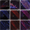
GFAP, AQP-4, SMA, and claudin-5 ex vivo immunohistochemistry. (A) GFAP staining reveals astrocytes entwining a diving arteriole, but no GFAP is visible along the larger pial surface artery. (B) AQP-4 staining reveals high levels of AQP-4 expression around capillaries and within the glia limitans. Although AQP-4 does not costain the inner SR101 sheath of the diving arteriole, it appears to be localized in the space occupied by the outer SR101 sheath (arrows). (C) SMA staining is visible outside SR101 staining that is colocalized with TIE2-GFP on this pial arteriole. (D to F) Pial surface artery showing fine structure of claudin-5 and SR101/Texas red hydrazide staining. A surface vein, with disordered claudin-5 staining, but no staining with SR101, is also visible. Arterial tight junctions outline elongated parallel structures running along the arteriole, whereas venous tight junctions have a less uniform arrangement. (G to I) Magnified region showing close apposition of claudin-5 expression and SR101 staining, suggesting that SR101 is localized within structures intimately associated with arteriolar endothelial tight junctions. AQP-4, aquaporin-4; GFAP, glial fibrillary acidic protein; GFP, green fluorescent protein; SMA, smooth muscle actin; SR101, sulforhodamine 101.

Comment in

References

    1. Altamura C, Dell'Acqua MI, Moessner R, Murphy DI, Lesch KP, Persico AM. Altered neocortical cell density and layer thickness in serotonin transporter knockout mice: a quantitation study. Cerebral Cortex. 2007;17:1394–1401. - PubMed
    1. Andresen J, Shafi NI, Bryan RM. Endothelial influences on cerebrovascular tone. J Appl Physiol. 2006;100:318–327. - PubMed
    1. Borowsky IW, Collins RC. Metabolic anatomy of brain: a comparison of regional capillary density, glucose metabolism, and enzyme activities. J Comp Neurol. 1989;288:401–413. - PubMed
    1. Bushong EA, Martone ME, Jones YZ, Ellisman MH. Protoplasmic astrocytes in CA1 stratum radiatum occupy separate anatomical domains. J Neurosci. 2002;22:183–192. - PMC - PubMed
    1. Cauli B, Ton XK, Rancillac A, Serluca N, Lambolez B, Rossier J, Hamel E. Cortical GABA interneurons in neurovascular coupling: relays for subcortical vasoactive pathways. J Neurosci. 2004;24:8940–8949. - PMC - PubMed

Publication types

LinkOut - more resources