

Gene expression analysis in SV-40 immortalized human corneal epithelial cells cultured with an air-liquid interface
- PMID: 21139686
- PMCID: PMC2994346
Gene expression analysis in SV-40 immortalized human corneal epithelial cells cultured with an air-liquid interface
Abstract
Purpose: To compare the global gene expression profile of stratified epithelia generated in vitro using simian virus 40 (SV40) immortalized human corneal epithelial cells with the previously reported gene expression of normal human corneal epithelia.
Methods: Immortalized cells expanded in submerged culture were grown in an air-liquid interface of liquid permeable collagen-coated filters to foster stratification and differentiation. Stratified epithelia displaying resistances exceeding 300 Ω·cm2 were dissolved in an RNA purification lysis buffer. Purified RNA was used to globally determine gene expression levels using high-density single-channel oligonucleotide microarrays. Raw hybridization readings were converted into relative gene expression levels using Robust Multi-array Average (RMA) algorithm. Expression levels for selected genes were validated by real-time RT-qPCR. The biologic significance of the gene expression profiles was interpreted with the help of several microarray software analysis tools and ad hoc thematical analysis.
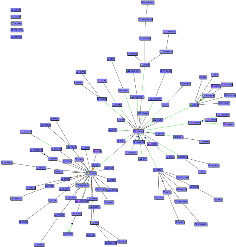
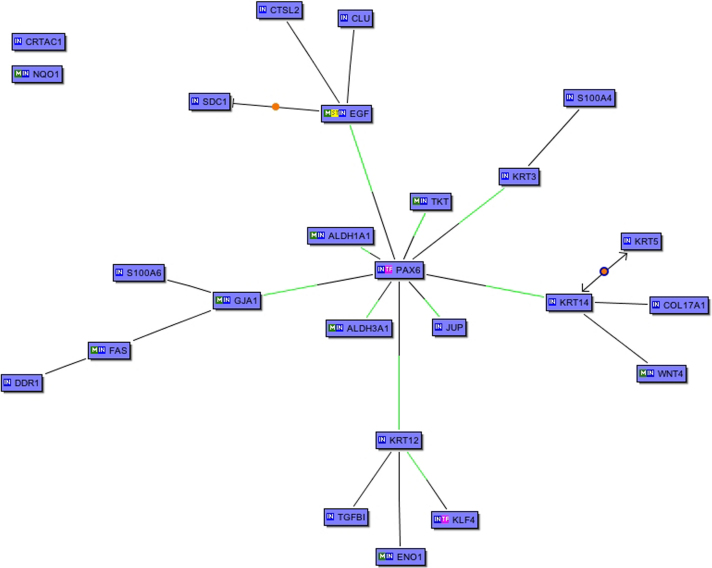
Results: The stratified cell culture to native epithelial comparison identified over- and under-expression in 22% and 14% of the probed genes, respectively. The larger expression decreases occurred in genes intimately associated with both the stratified epithelial lineage at large such as keratin 14 and the corneal phenotype, such as keratin 12, connexin 43, aldehyde dehydrogenases (ALDHs), and paired box gene 6 (PAX6) and its whole downstream transcriptome. Overexpression related to genes associated with cell cycling stimulation.
Conclusions: The results indicate that the stratified corneal epithelial cell model generated using SV40 immortalized cells may be useful only in certain research applications. Extrapolations of studies with these cells to actual tissue cells should be done with a great deal of caution.
Figures
References
-
- Wolosin JM. Regeneration of resistance and ion transport in rabbit corneal epithelium after induced surface cell exfoliation. J Membr Biol. 1988;104:45–55. - PubMed
-
- Sokol JL, Masur SK, Asbell PA, Wolosin JM. Layer-by-layer desquamation of corneal epithelium and maturation of tear-facing membranes. Invest Ophthalmol Vis Sci. 1990;31:294–304. - PubMed
-
- Wang Y, Chen M, Wolosin JM. ZO-1 in corneal epithelium; stratal distribution and synthesis induction by outer cell removal. Exp Eye Res. 1993;57:283–92. - PubMed
-
- Wolosin JM, Chen M. Ontogeny of corneal epithelial tight junctions: stratal locale of biosynthetic activities. Invest Ophthalmol Vis Sci. 1993;34:2655–64. - PubMed
Publication types
MeSH terms
Substances
Grants and funding
LinkOut - more resources
Full Text Sources
Other Literature Sources
Molecular Biology Databases