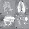
Decision-making in burst fractures of the thoracolumbar and lumbar spine
- PMID: 21139777
- PMCID: PMC2989512
- DOI: 10.4103/0019-5413.36986
Decision-making in burst fractures of the thoracolumbar and lumbar spine
Abstract
The most common site of injury to the spine is the thoracolumbar junction which is the mechanical transition junction between the rigid thoracic and the more flexible lumbar spine. The lumbar spine is another site which is more prone to injury. Absence of stabilizing articulations with the ribs, lordotic posture and more sagitally oriented facet joints are the most obvious explanations. Burst fractures of the spine account for 14% of all spinal injuries. Though common, thoracolumbar and lumbar burst fractures present a number of important treatment challenges. There has been substantial controversy related to the indications for nonoperative or operative management of these fractures. Disagreement also exists regarding the choice of the surgical approach. A large number of thoracolumbar and lumbar fractures can be treated conservatively while some fractures require surgery. Selecting an appropriate surgical option requires an in-depth understanding of the different methods of decompression, stabilization and/or fusion. Anterior surgery has the advantage of the greatest degree of canal decompression and offers the benefit of limiting the number of motion segments fused. These advantages come at the added cost of increased time for the surgery and the related morbidity of the surgical approach. Posterior surgery enjoys the advantage of being more familiar to the operating surgeons and can be an effective approach. However, the limitations of this approach include inadequate decompression, recurrence of the deformity and implant failure. Though many of the principles are the same, the treatment of low lumbar burst fractures requires some additional consideration due to the difficulty of approaching this region anteriorly. Avoiding complications of these surgeries are another important aspect and can be achieved by following an algorithmic approach to patient assessment, proper radiological examination and precision in decision-making regarding management. A detailed understanding of the mechanism of injury and their unique biomechanical propensities following various forms of treatment can help the spinal surgeon manage such patients effectively and prevent devastating complications.
Keywords: Burst fracture; lumbar fracture; thoracolumbar fracture.
Conflict of interest statement
Figures


References
-
- Melton LJ, 3rd, Thamer M, Ray NF, Chan JK, Chesnut CH, 3rd, Einhorn TA, et al. Fractures attributable to osteoporosis: Report from the National Osteoporosis Foundation. J Bone Miner Res. 1997;12:16–23. - PubMed
-
- Hu R, Mustard CA, Burns C. Epidemiology of incident spinal fracture in a complete population. Spine. 1996;21:492–9. - PubMed
-
- Flanders AE. Thoracolumbar trauma imaging overview. Instructional Course Lectures. 1999;48:429–31. - PubMed
-
- Holdsworth FW. Fractures, dislocations and fracture/dislocation of the spine. J Bone Joint Surg Br. 1963;45:6–20.
-
- Bensch FV, Koivikko MP, Kiuru MJ, Koskinen SK. The incidence and distribution of burst fractures. Emerg Radiol. 2006;12:124–9. - PubMed
LinkOut - more resources
Full Text Sources
