

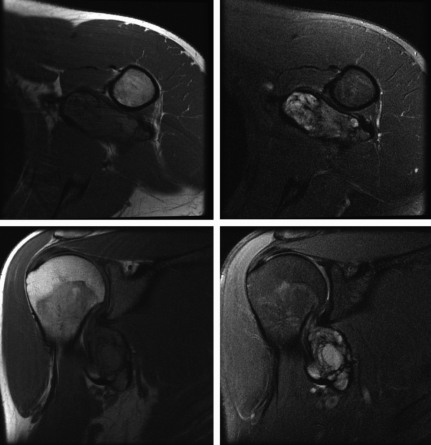
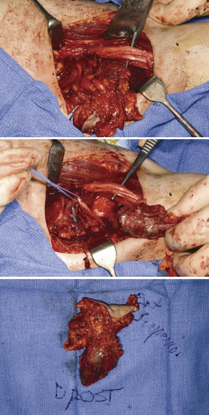
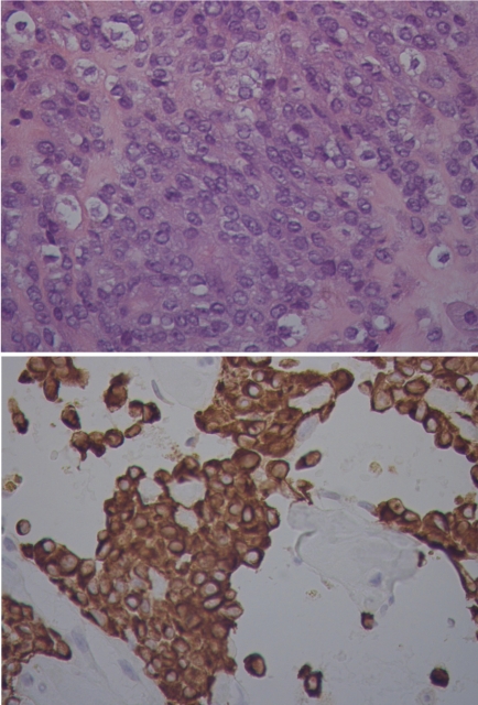
Angiomatoid fibrous histiocytoma in a 25-year-old male
- PMID: 21139823
- PMCID: PMC2994511
- DOI: 10.4081/rt.2010.e20
Angiomatoid fibrous histiocytoma in a 25-year-old male
Abstract
Angiomatoid fibrous histiocytoma (AFH) is a rare disease that is often misdiagnosed initially. Patients can present with a clinical picture concerning for other diseases, and pathologic review is not always revealing. Molecular diagnostics are increasingly being utilized to detect gene fusions characteristic for AFH. Surgery remains the mainstay of management, and can effectively control local recurrences and metastases. Herein we describe a case report of a 25-year-old gentleman whose presentation was concerning for lymphoma. Subsequently we review of the relevant literature.
Keywords: angiomatoid fibrous histiocytoma; soft tissue tumor..
Conflict of interest statement
Conflict of interest: the authors report no conflicts of interest.
Figures
References
-
- Enzinger FM. Angiomatoid malignant fibrous histiocytoma: a distinct fibrohistiocytic tumor of children and young adults simulating a vascular neoplasm. Cancer. 1979;44:2147–57. - PubMed
-
- Fanburg-Smith JC, Miettinen M. Angiomatoid “malignant” fibrous histiocytoma: a clinicopathologic study of 158 cases and further exploration of the myoid phenotype. Hum Pathol. 1999;30:1336–43. - PubMed
-
- Costa MJ, Weiss SW. Angiomatoid malignant fibrous histiocytoma. A follow-up study of 108 cases with evaluation of possible histologic predictors of outcome. Am J Surg Pathol. 1990;14:1126–32. - PubMed
-
- Hasegawa T, Seki K, Ono K, Hirohashi S. Angiomatoid (malignant) fibrous histiocytoma: a peculiar low-grade tumor showing immunophenotypic heterogeneity and ultrastructural variations. Pathol Int. 2000;50:731–8. - PubMed
-
- De Beuckeleer L. Fibrohistiocytic tumors. In: De Schepper AM, Parizel PM, De Beuckeleer L, Vanhoenacker F, editors. Imaging of soft tissue tumors. Berlin, Heidelberg, New York: Springer; 2001. pp. 181–93.
Publication types
LinkOut - more resources
Full Text Sources
